http://www.uni-mainz.de/ FB/Medizin/Anatomie/workshop
This was presented on May, 25th on the 6th International Symposium of Clinical and Applied Anatomy, Graz, Austria.
|
http://www.uni-mainz.de/ FB/Medizin/Anatomie/workshop This was presented on May, 25th on the 6th International Symposium of Clinical and Applied Anatomy, Graz, Austria. |
|
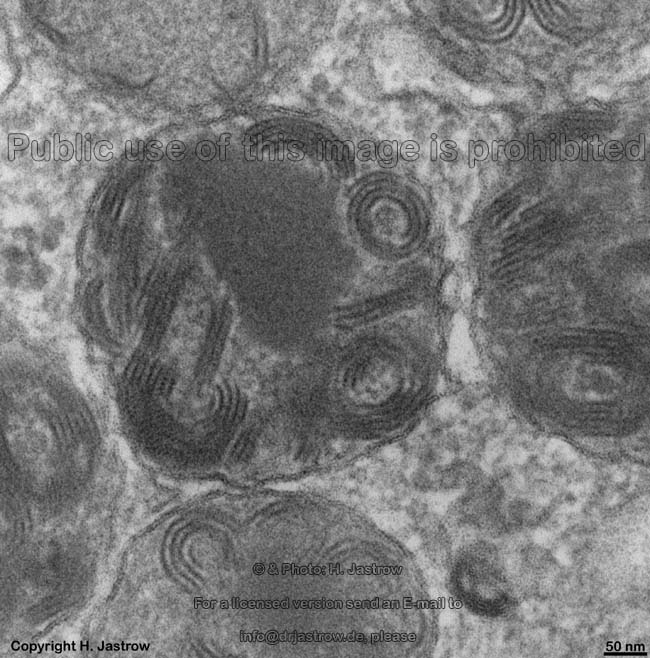
Fig.1
 |
Dear colleagues,
the internet became the most important source of information today thus there are plenty offers concerning anatomy. However, most of them cover only small parts of the entire field. The workshop Anatomy for the internet of which you see the homepage in Fig.2 has been established to present interlinked |
Fig.2 - original
WAI page
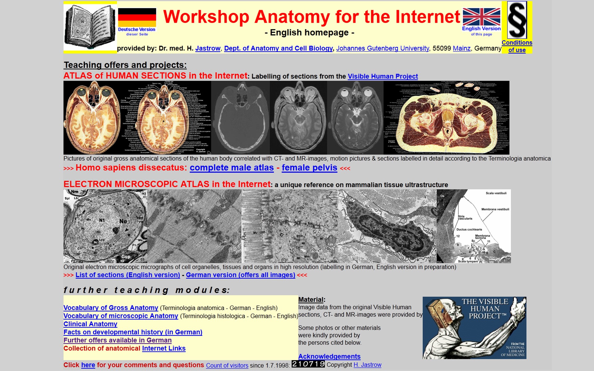 |
Fig.3
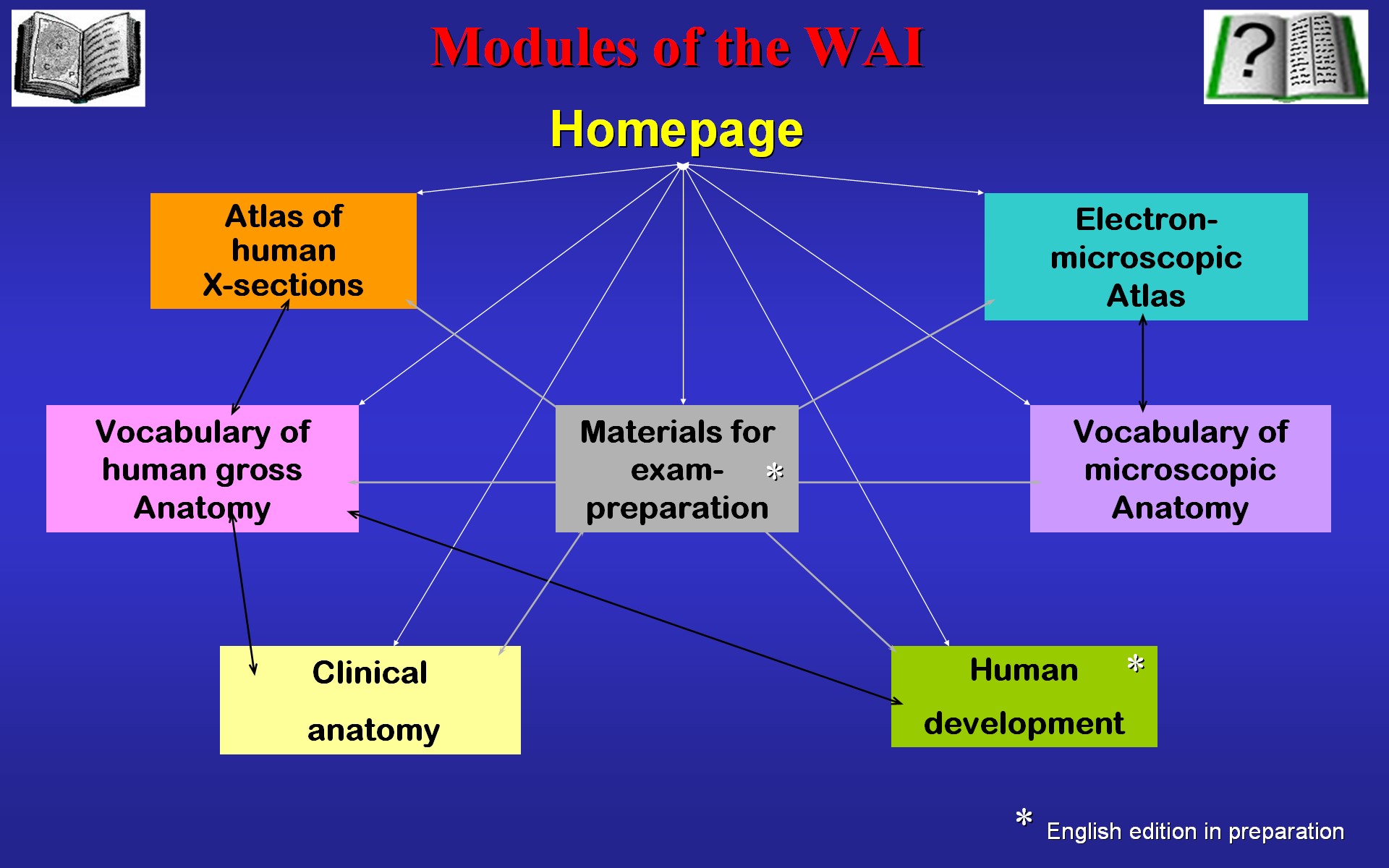 |
anatomy web-teaching modules. These
modules are linked to each other as shown in Fig.3 to enable the
user to gain a comprehensive picture of the human body. The main modules
are an electron microscopic atlas and a complete atlas of human cross sections.
These atlases both are linked to vocabularies explaining applied terms
also providing background information. Some materials of clinical anatomy
as well as a complete overview of human development are provided with some
special texts and topics for exam preparation of which an English version
is in preparation. Lets have a first glance at the atlas of human cross
sections
It contains all sections of the visible human male which can be reached from the index page shown in Fig.4 providing links to overview pages of 35 segments. The atlas is language independent as far as possible but handling is also explained in four languages. |
Fig.4 - original
WAI page
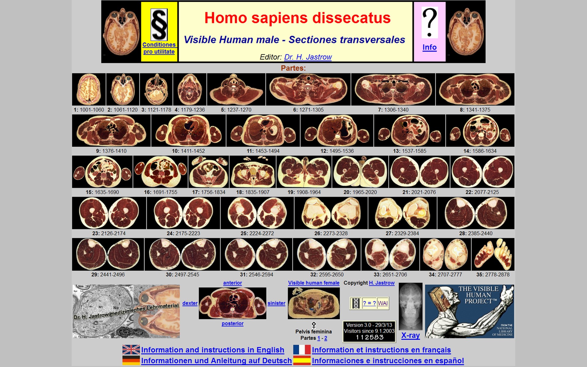 |
Fig.5 - original WAI page 1
- 2
 |
As explained in Fig.5, navigation bars
with small symbols allow to reach any desired topic quickly to enable easy
international use by anybody interested in human cross-sections.
The atlas is based on the data of the visible human project which were processed as noted in Fig.6. |
Fig.6 - original
WAI page
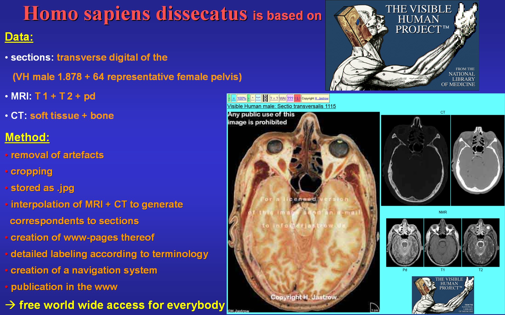 |
Fig.7 - original
WAI page
 |
Fig.7 shows an example of a section overview page. If we chose 1878 here by clicking the page of the appropriate page of the pelvis appears with corresponding radiological images (Fig.8). If we want to follow the spermatic cord a little, we can click on the next section symbol. | Fig.8 - original
WAI page
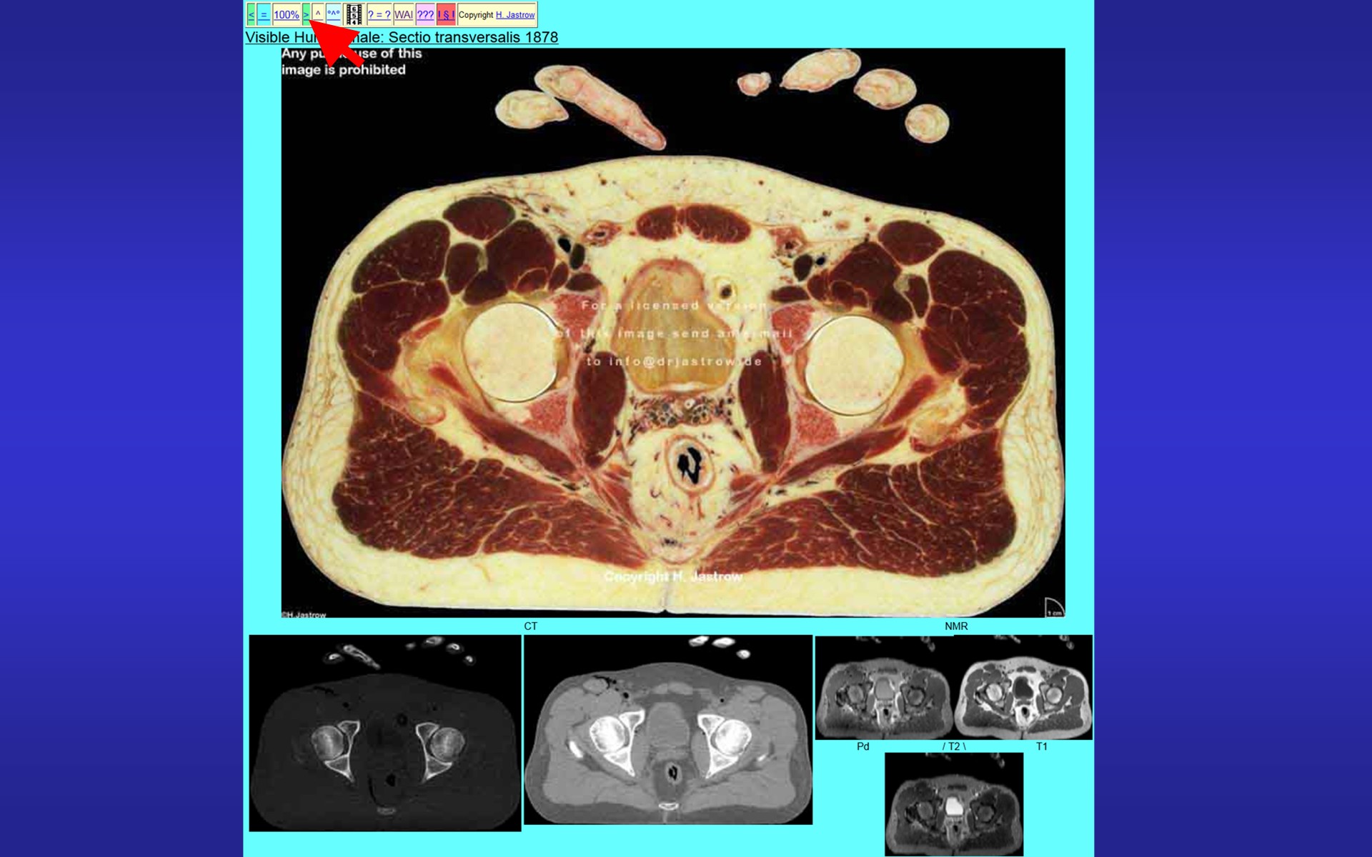 |
Fig.9 - original
WAI page
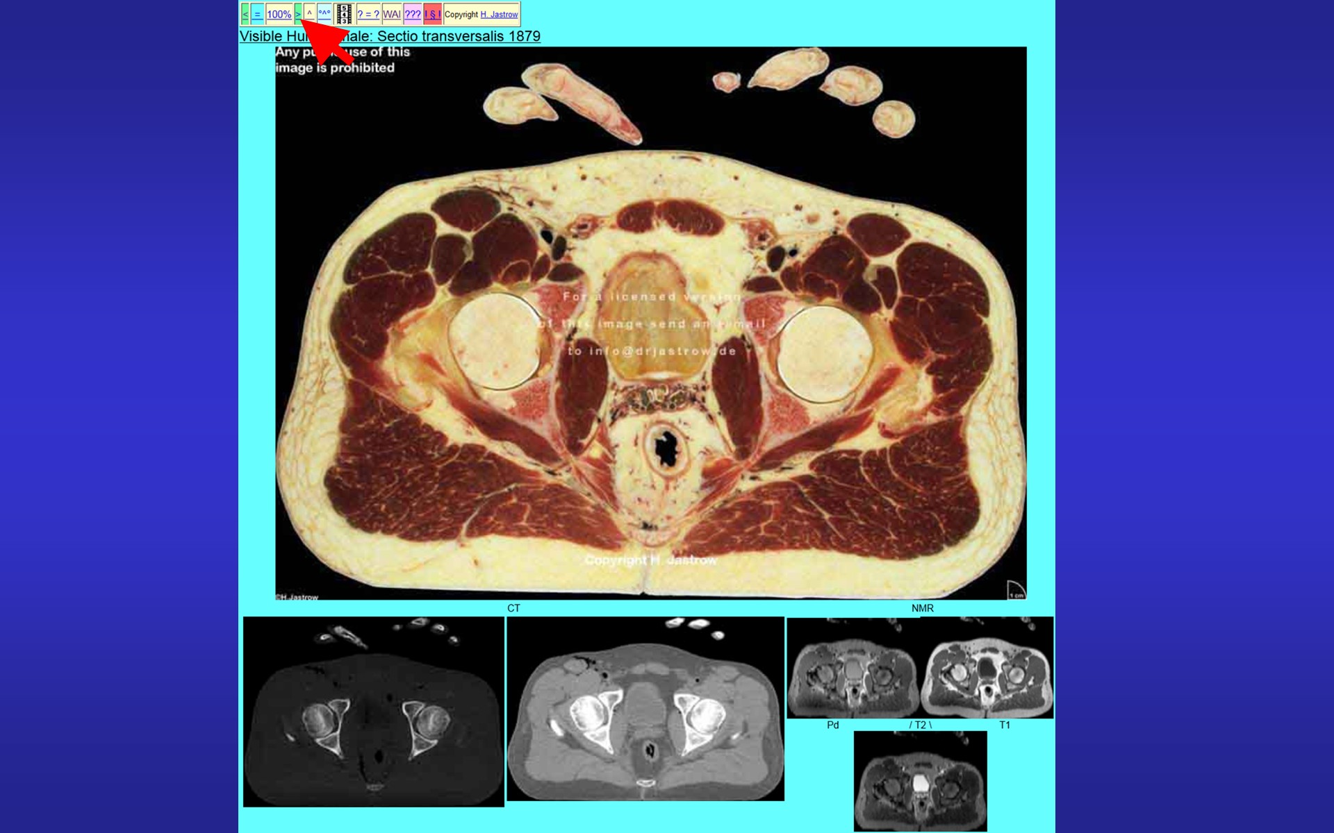 |
Now the next section appears (Fig.9) which is 1mm below the first one. By a further click we again get 1 mm deeper (Fig.10) | Fig.10 - original
WAI page
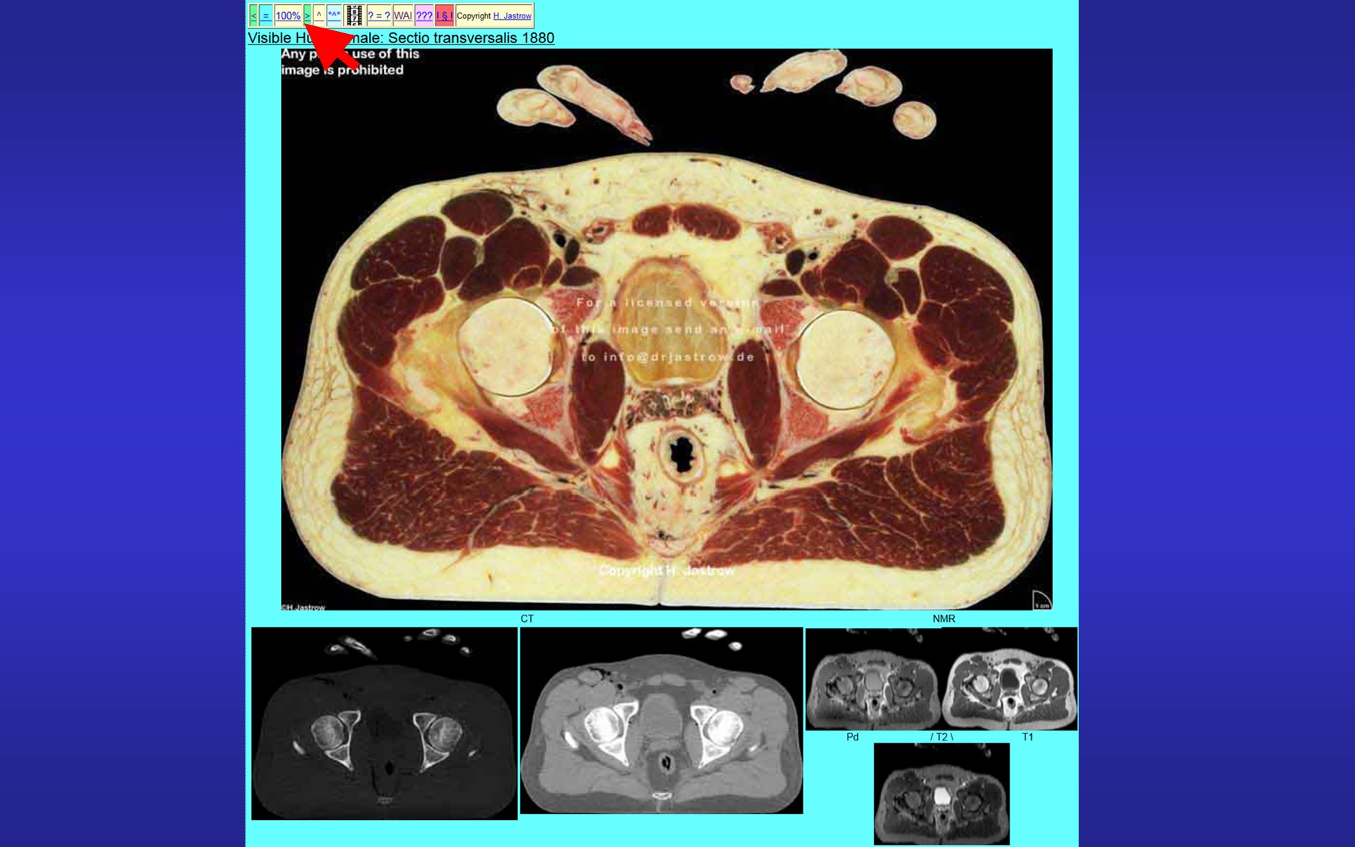 |
Fig.11 - original
WAI page
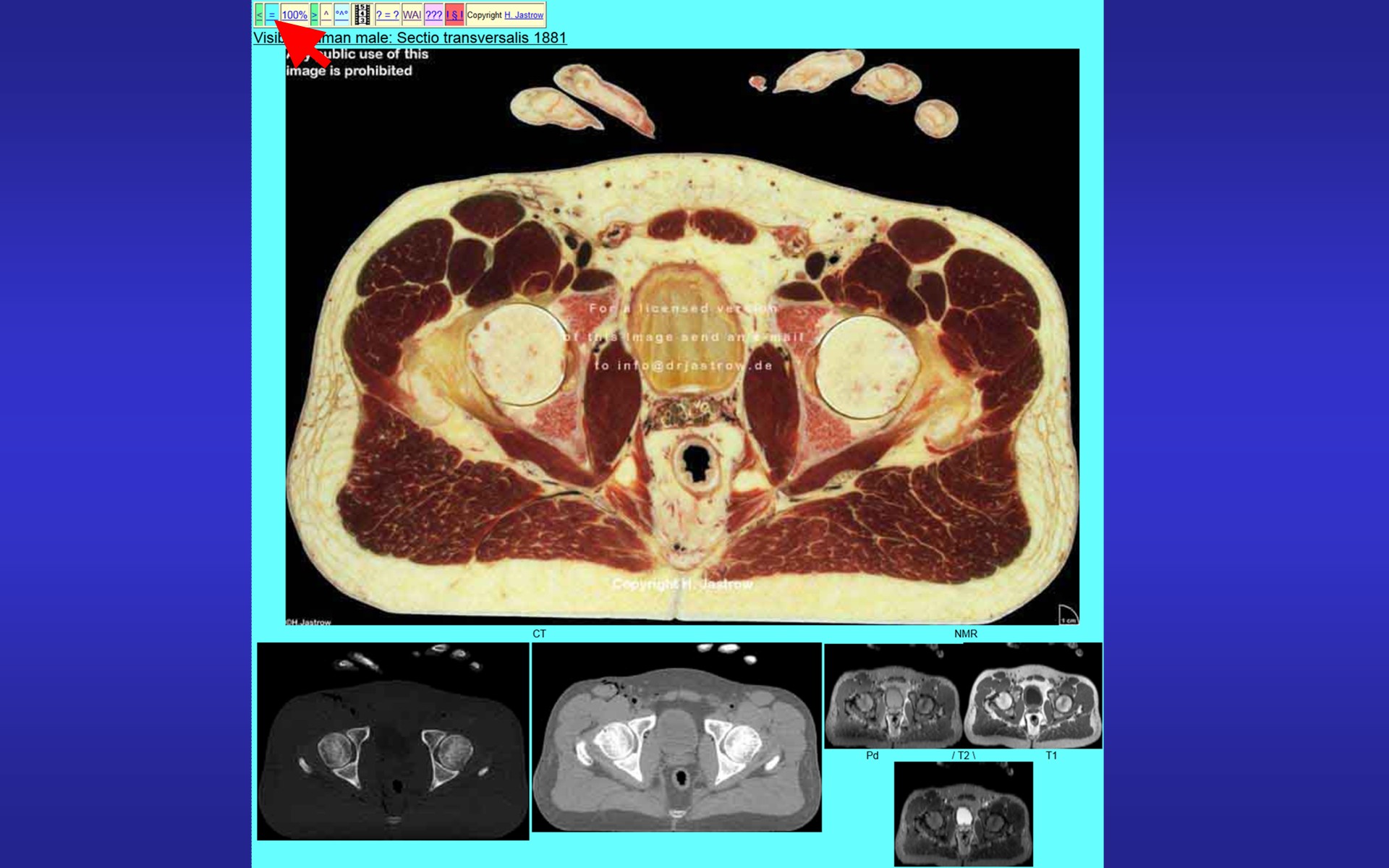 |
in the section series. A further click allows a follow up on the adjacent section (Fig.11). When we now want to see which structures are visible on the section we can click on the symbol for labelled section to see Fig.12 which shows a detailed labelling including the structures of the spermatic cord. However, an animation through serial sections will surely be able to demonstrate the course of the spermatic cord even better. A click here | Fig.12 - original
WAI page
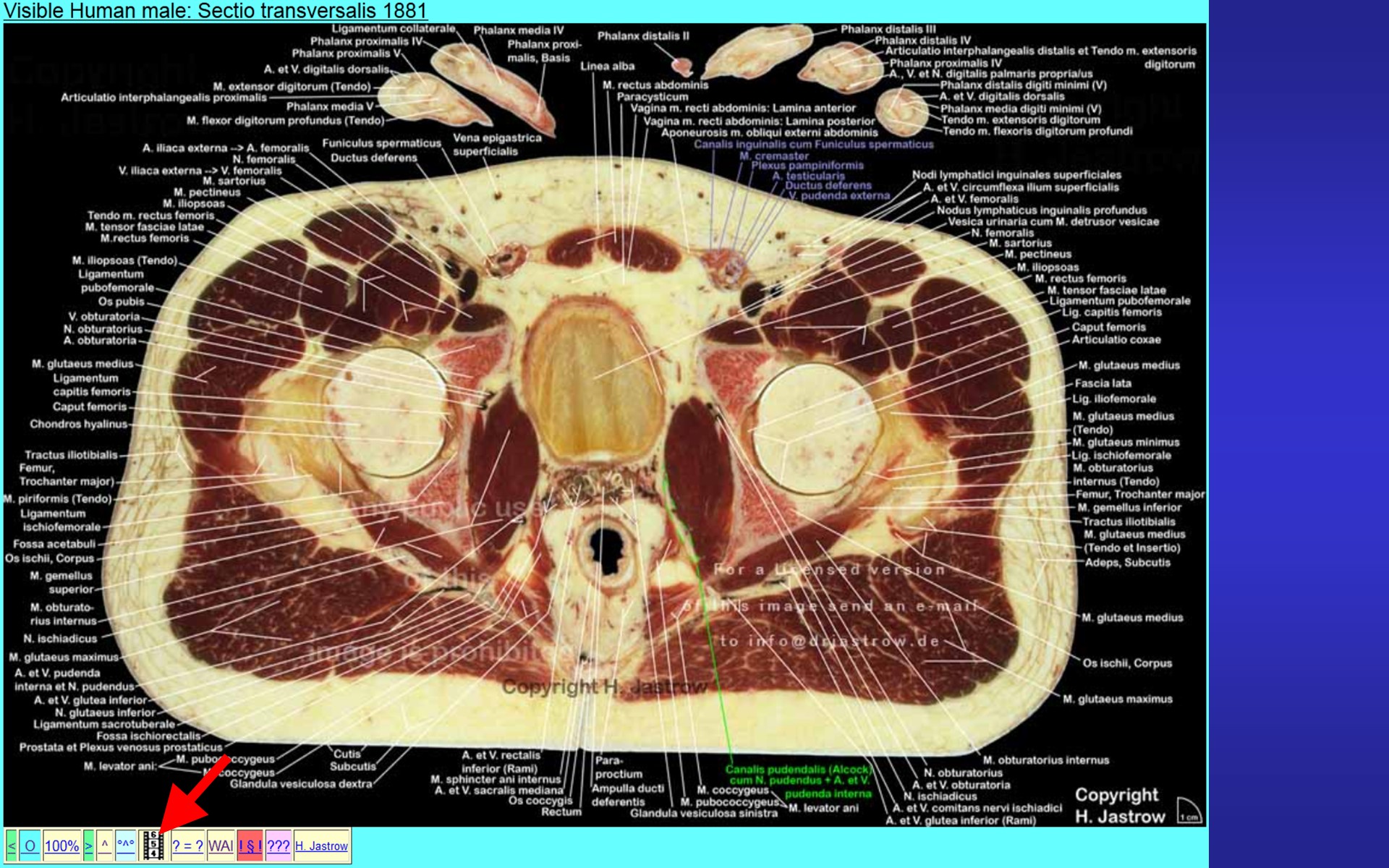 |
Fig.13 - original
WAI page
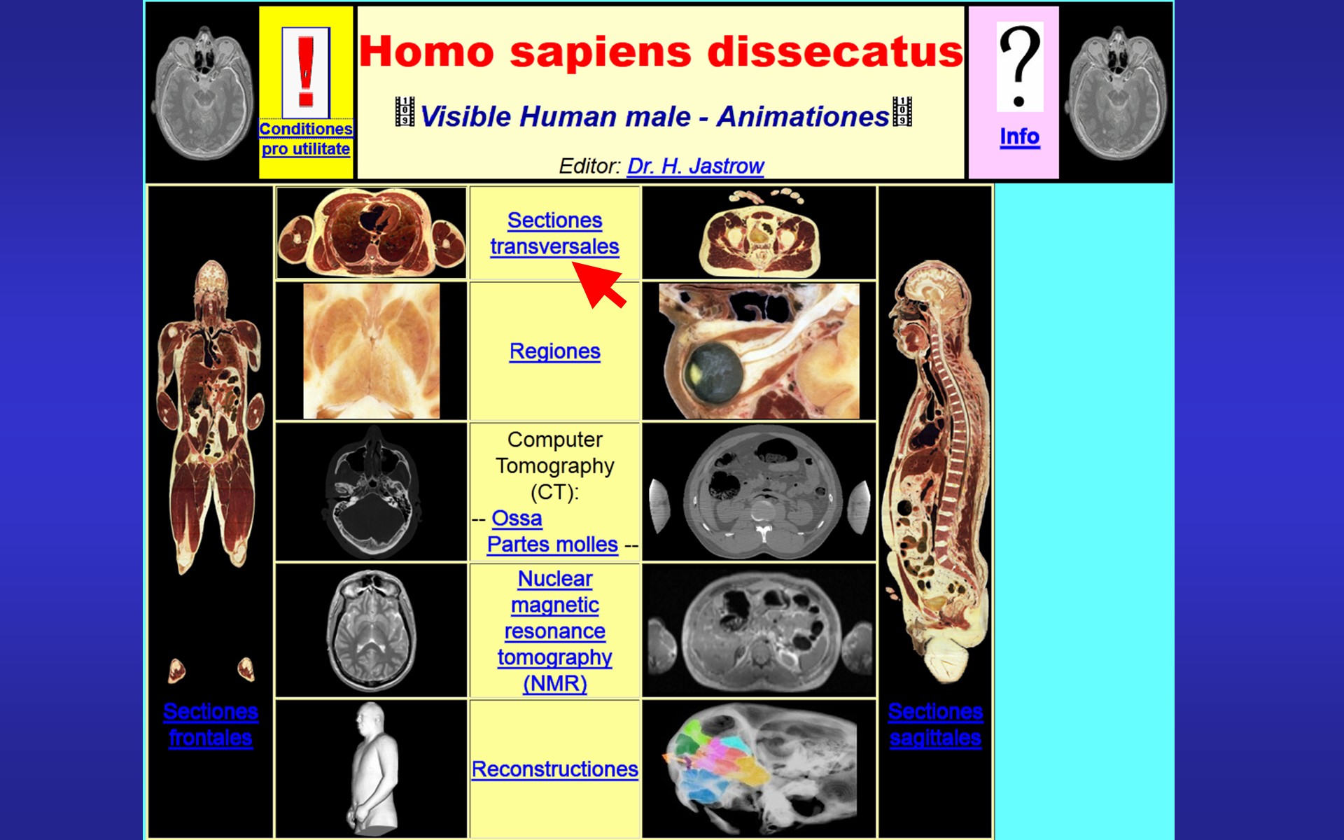 |
Calls up the overview page with animations of the visible human male data set (Fig.13) from which we choose the "sectiones transversales" leading to a page (Fig.14) offering an animation through the sections of the pelvis. A click here | Fig.14 - original
WAI page
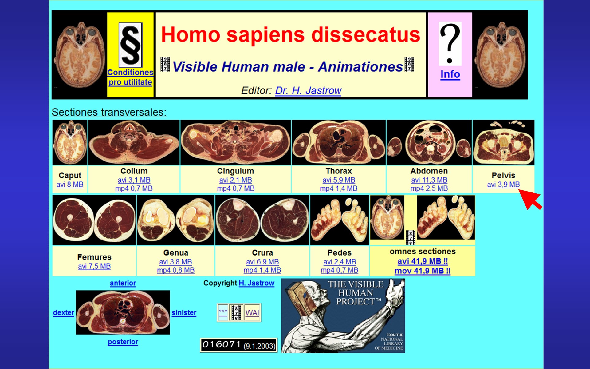 |
Fig.15
 |
opens a motion picture of all sections of the pelvis (Fig.15) where we can follow the course of the spermatic cord and explore its topography. Other animations show serial bone CTs (Fig.16) | Fig.16 - original
WAI page
 |
Fig.17 - original
WAI page
 |
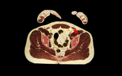
or soft tissue CT scan animations (Fig. 17). Further, some animations of MR-image sequences are available (Fig. 18). | Fig.18 - original
WAI page
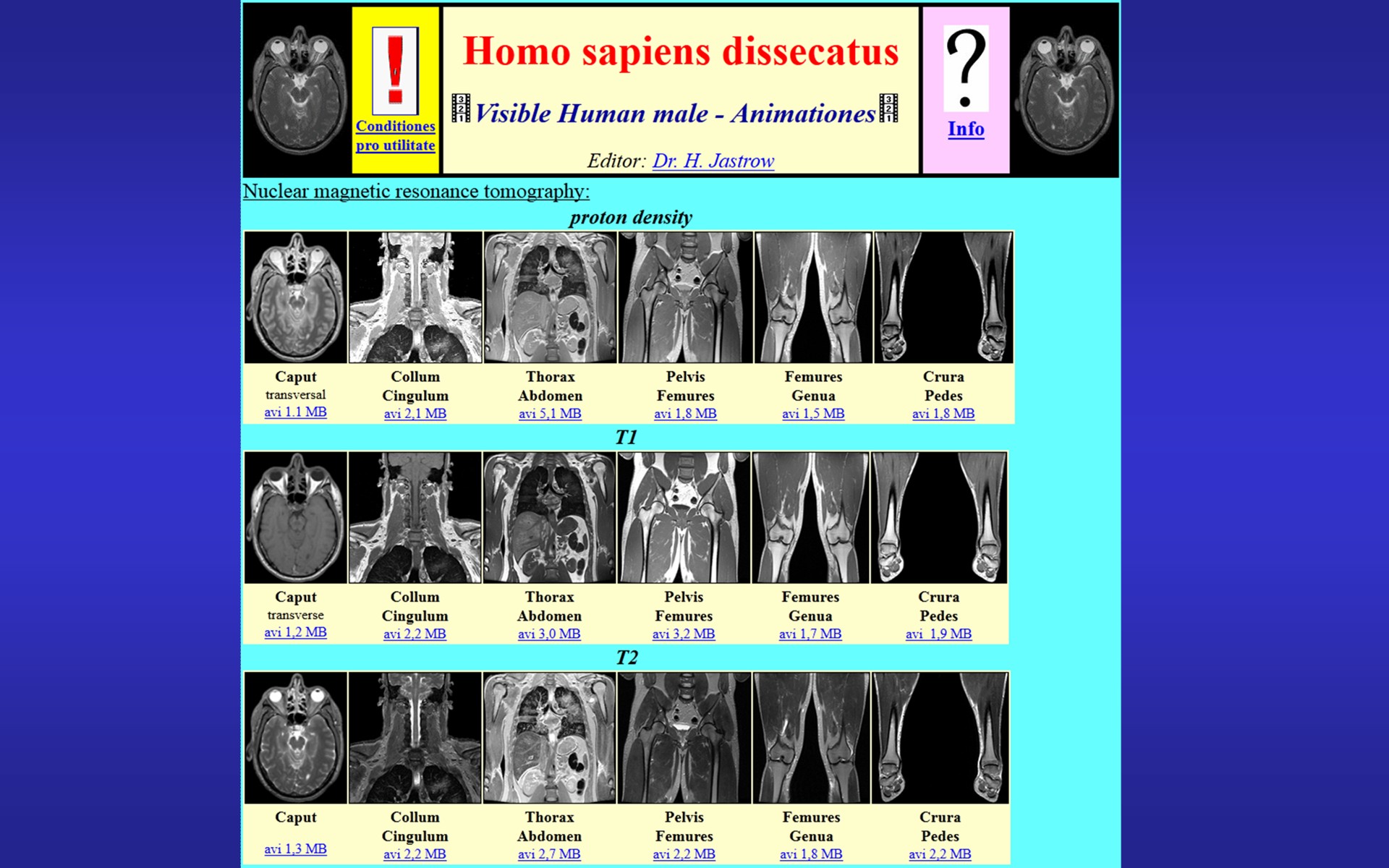 |
Fig.19 - original
WAI page
 |
Now lets have a look on the clinical anatomy page (Fig.19) which offers some animations of swinging vocal plicas or a stereoscopic image of the optic nerve papilla (Fig.20). From here we can go with a link to the | Fig.20 - original WAI page 1
- 2
 |
Fig.21 - original
WAI page
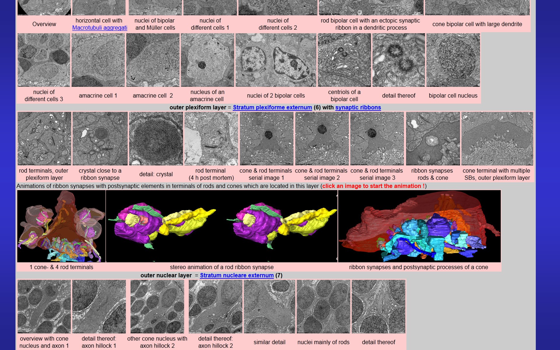 |
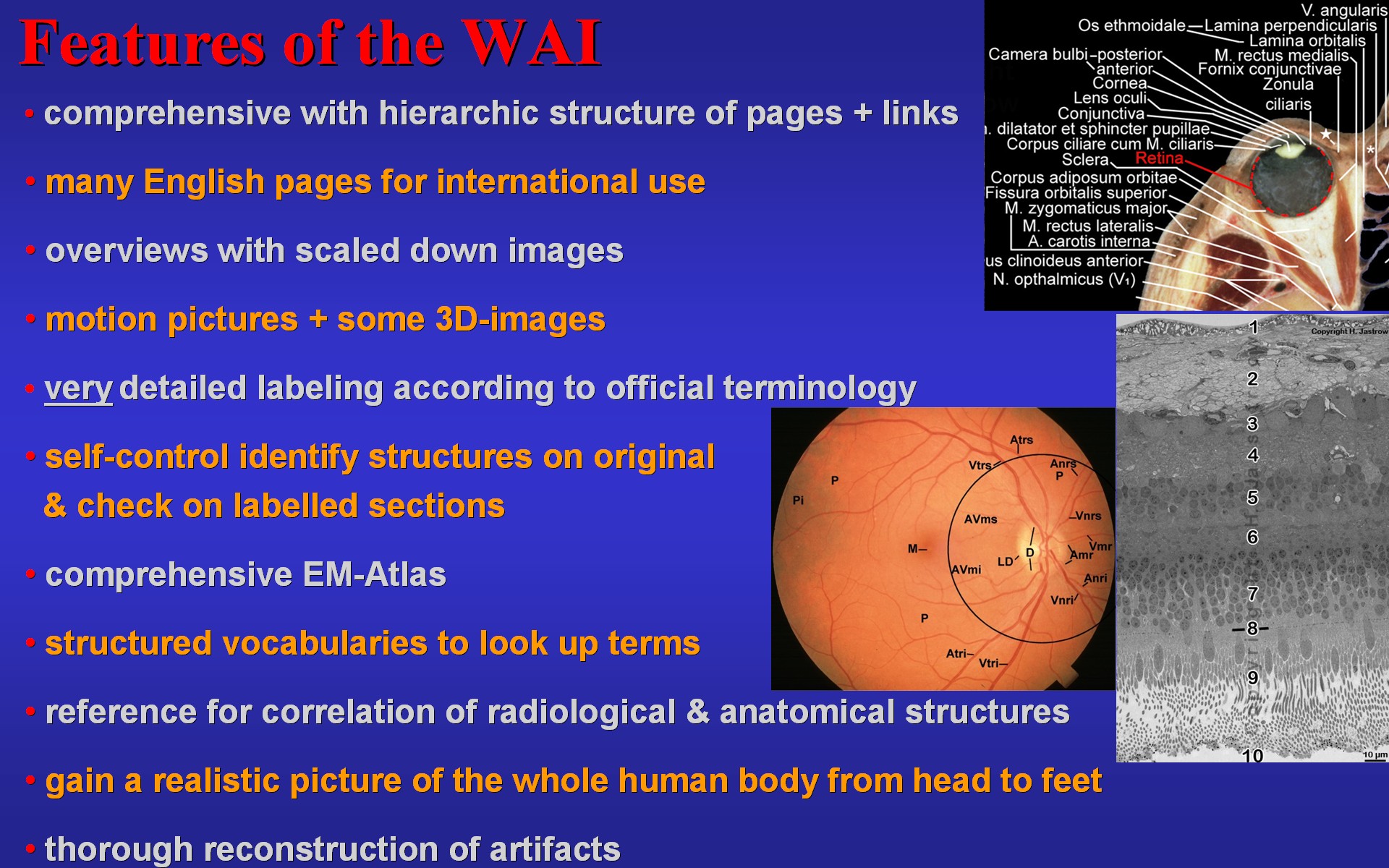
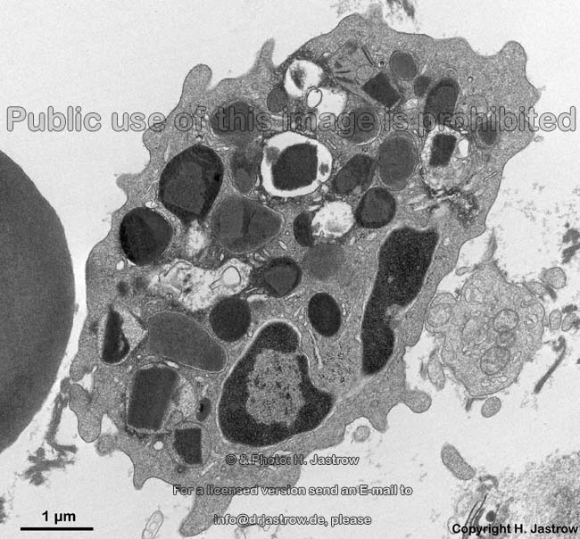
human retina page of the electron microscopic atlas (part thereof shown in Fig.21) which offers some stereo animations of reconstructed ribbon synapses. The electron microscopic atlas is based on over 1,500 mainly transmission electron microscopic images. From the index page (left in Fig.22) overview pages with minimized images and explaining text can be retrieved. | Fig.22 - original
WAI page
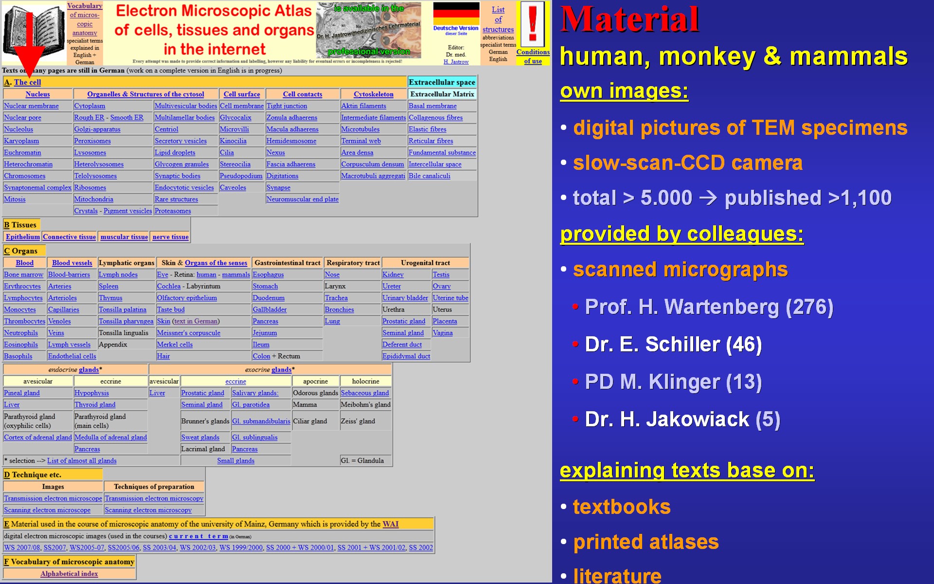 |
Fig.23 - original
WAI page
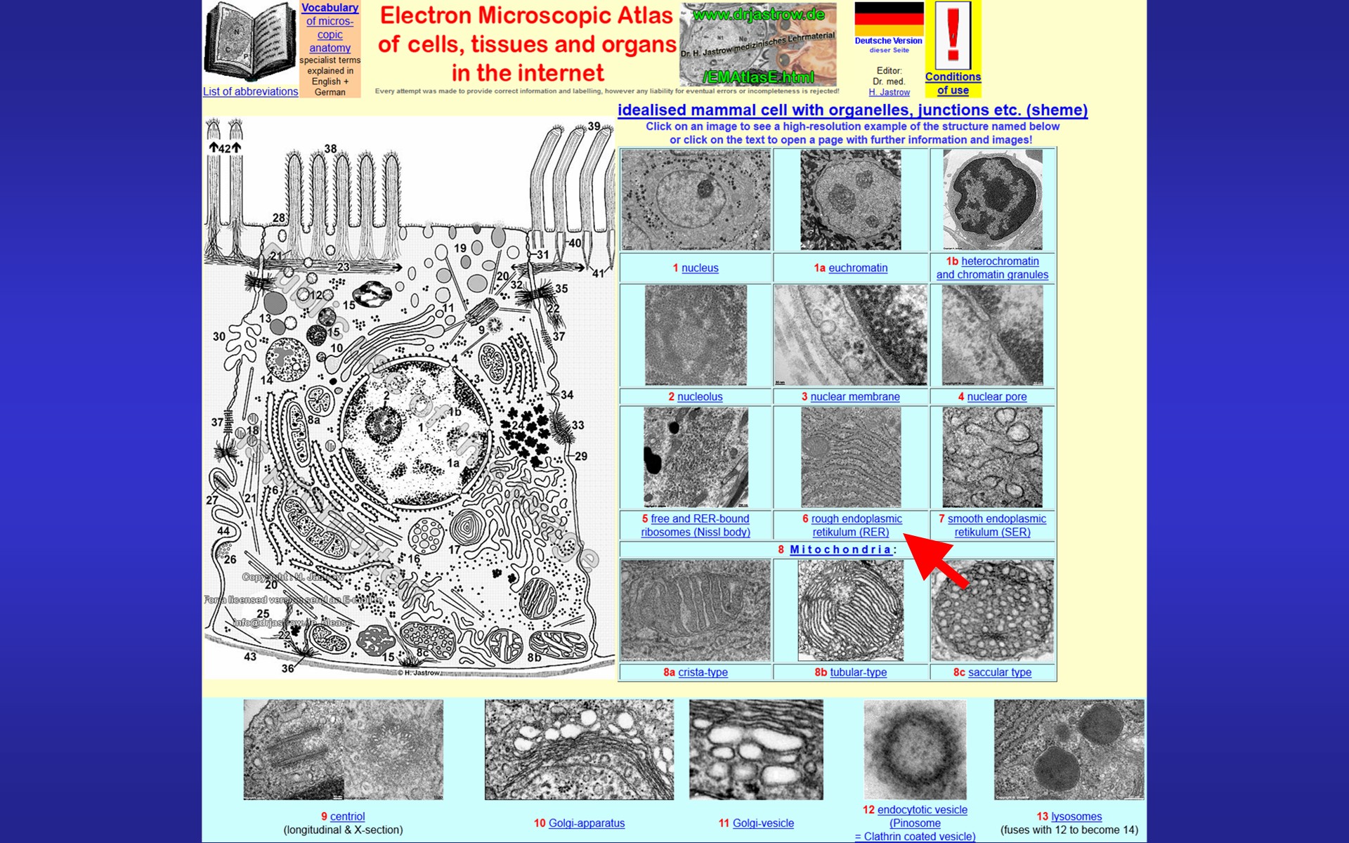 |
One of them is the page shown in Fig.23 with a schematic drawing of a cell demonstrating organelles of which miniatures are linked to overview pages. When we click on the rough endoplasmic reticulum the appropriate page providing small overview images as well a text on RER is called (Fig.24). | Fig.24 - original
WAI page
 |
Fig.25 - original
WAI page
 |
Back to the index page (Fig.25) we now have a look at the muscular tissue and click here loads a page of differential diagnosis of muscle tissues with small sample images (Fig.26). If we click on the picture for skeletal muscle | Fig.26 - original
WAI page
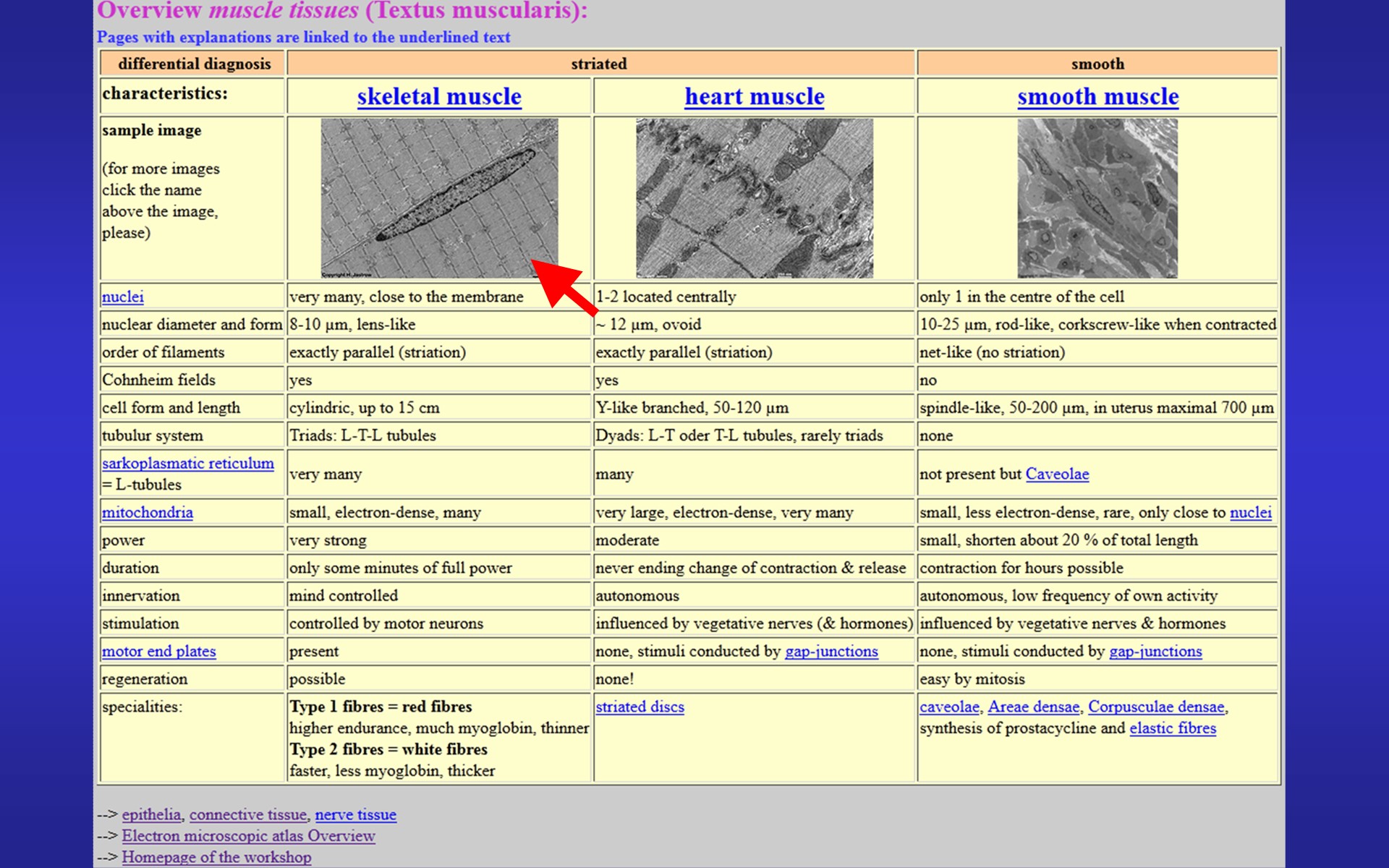 |
Fig.27 - original
WAI page
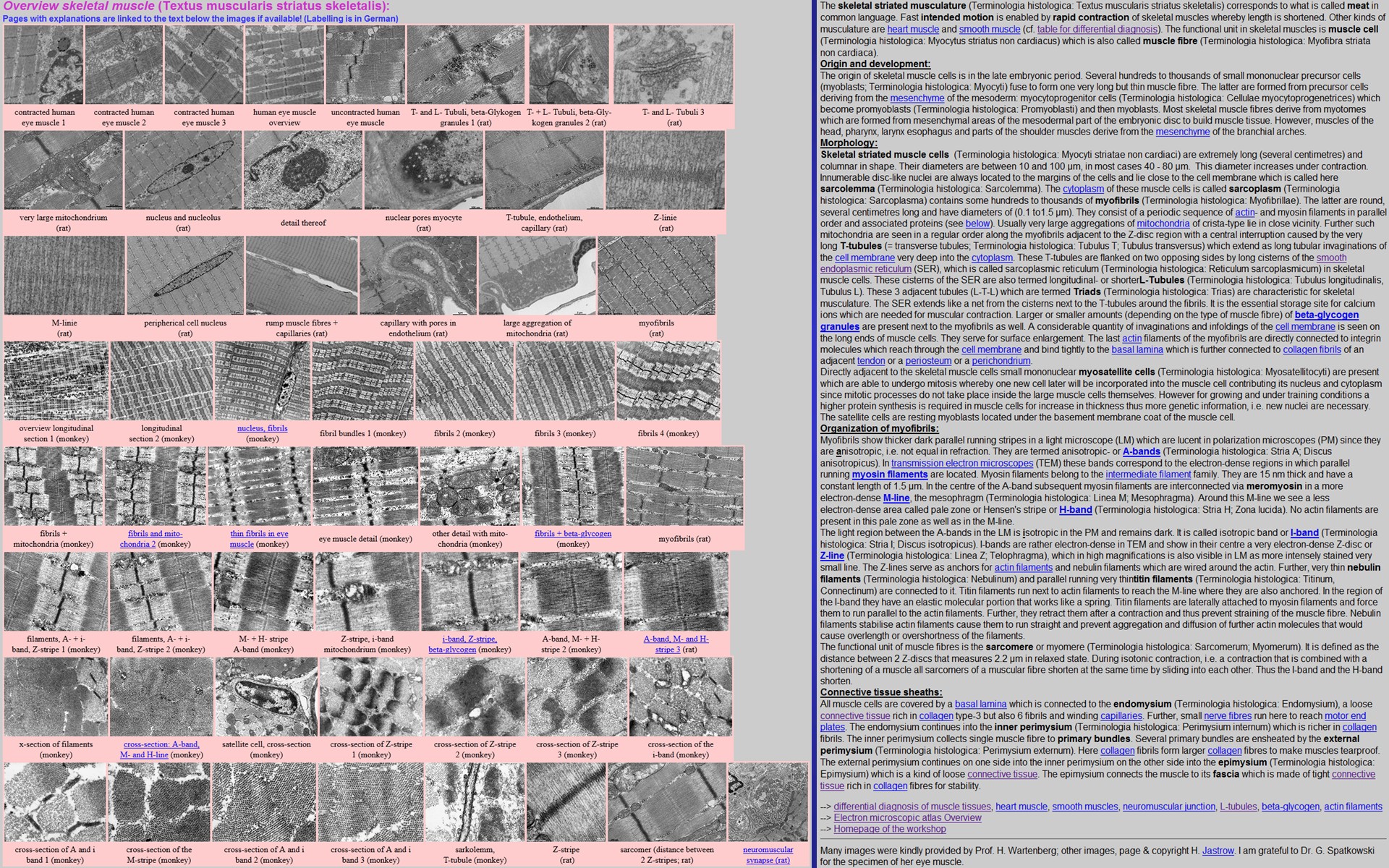 |
the skeletal muscle page appears (Fig.27).
Besides a comprehensive text links are provided to over 55 original images
and 8 blue underlined labelled images.
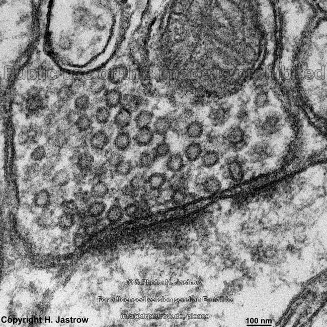
Now we want to look up "desmosome" in the vocabulary of microscopic anatomy. Thus we click on D to find a short description for more images and information (Fig.28) we click here |
Fig.28 - original WAI page 1
- 2
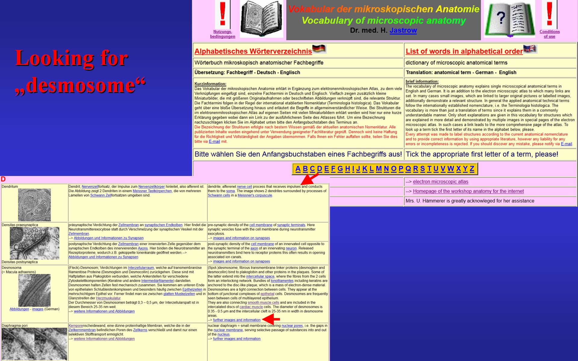 |
Fig.29 - original
WAI page
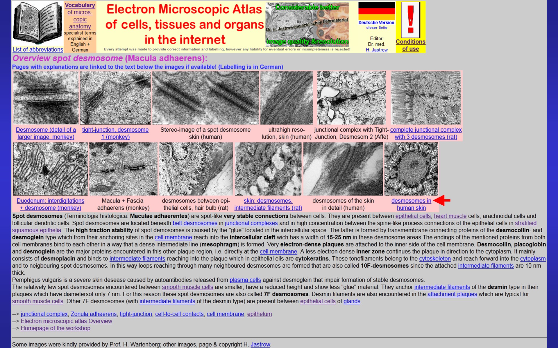 |
to see the EM atlas page of spot desmosomes (Fig.29). A click on the linked text under the image from the skin leads to the page with the labelled and explained image (Fig.30). All pages are available in German. However, many of them are already also provided in English. At present, over 200 of most interesting images are labelled. Besides background information about histology, physiology and biochemistry of described structures we find information on electron microscopes and specimen preparation in the EM-atlas. | Fig.30 - original
WAI page
 |
Fig.31 - original WAI page 1
- 2
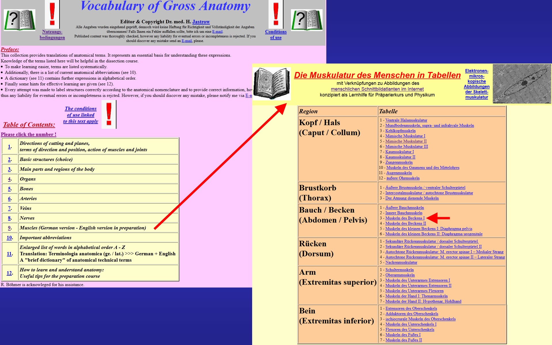 |
Now lets explore one of the most viewed pages of the vocabulary of macroscopic anatomy which is the overview page of muscles. It can be retrieved from an overview page (Fig.31, left) also providing links to short summary pages on arteries, veins and other topics. When we choose muscles (Fig.31, right) and then specify muscles of the pelvis the page shown in Fig. 32 comes up that lists muscle group & -name, origin, insertion, innervation and function of pelvic muscles. Further, as we can see here on the example of the obturator externus, a section of the cross-sectional atlas is suggested to | Fig.32 - original
WAI page
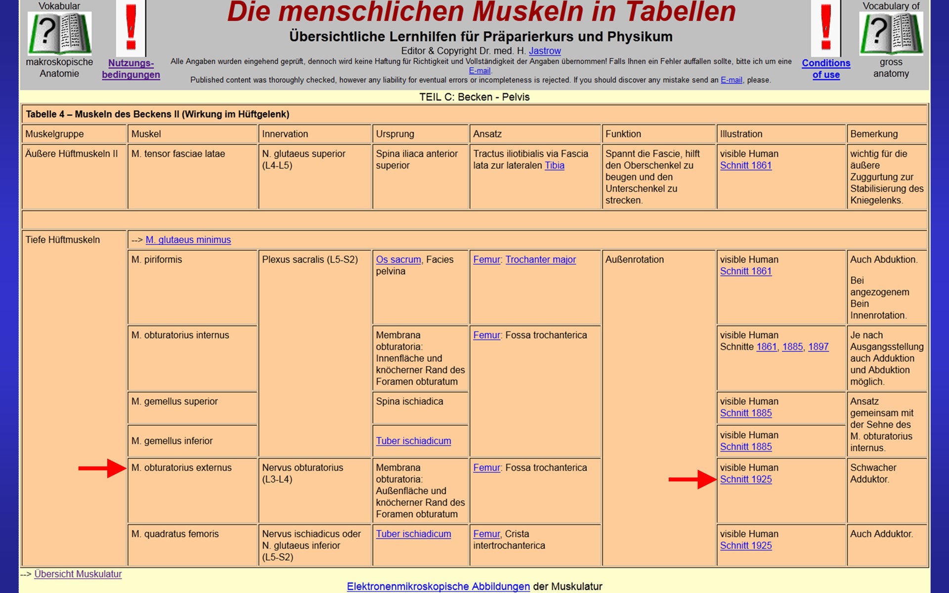 |
Fig.33 - original
WAI page
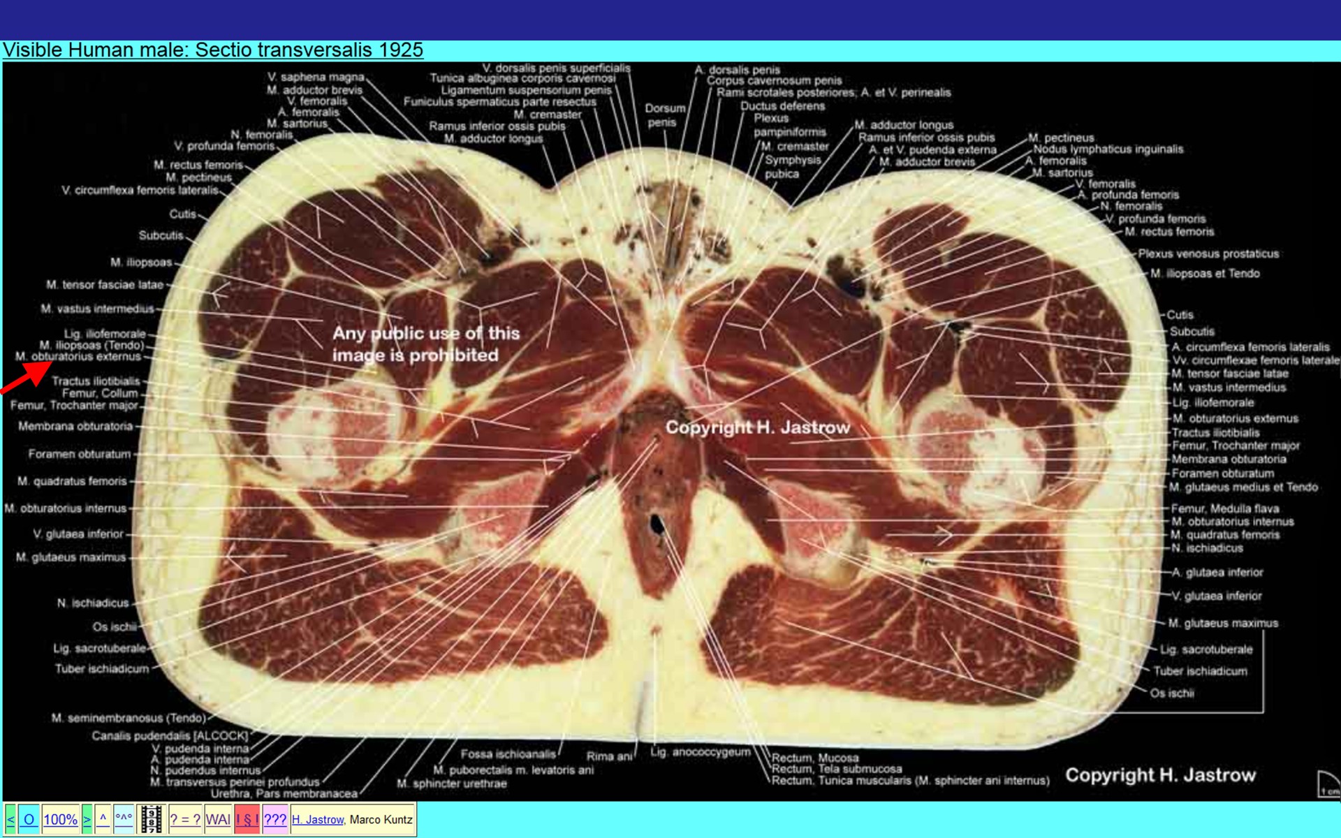 |
view the muscle besides many other labelled structures (Fig.33).
Facts & data on human development are available for each day of the embryonic to early foetal period and then in a weekly survey in German (Fig.34). An example can be seen on the right. One can march from one to the next day to follow the development of organs in a chronological manner. |
Fig.34 - original WAI pages 1
- 2
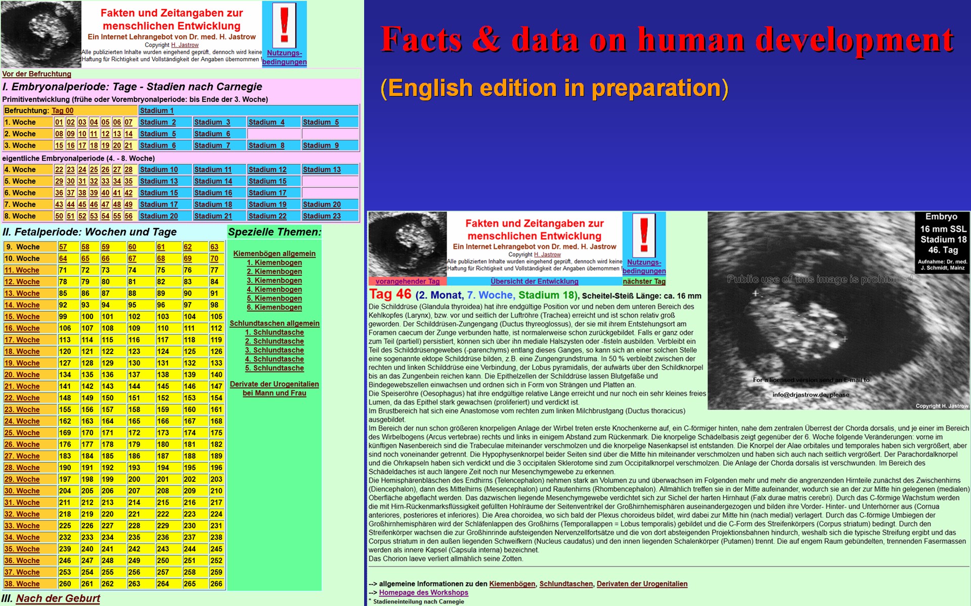 |
Fig.35 - original
WAI page
 |
There are some pages of themes of special interest for exam preparation accessible from the page shown in Fig.35. It, e.g., provides links to the extensively labelled section of the spinal cord with explaining text demonstrated in Fig. 36, | Fig.36 - original
WAI page
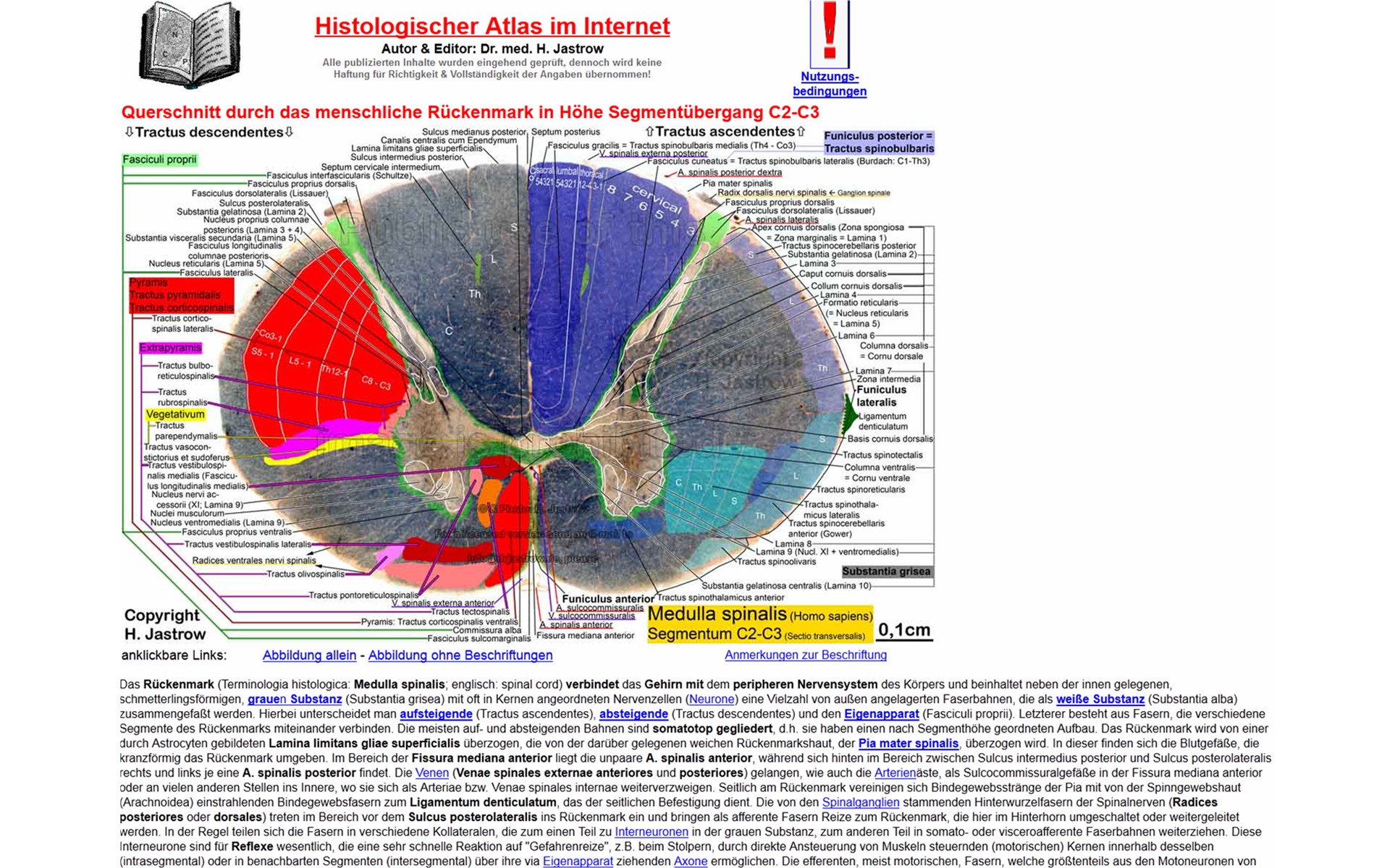 |
Fig.37 - original
WAI page
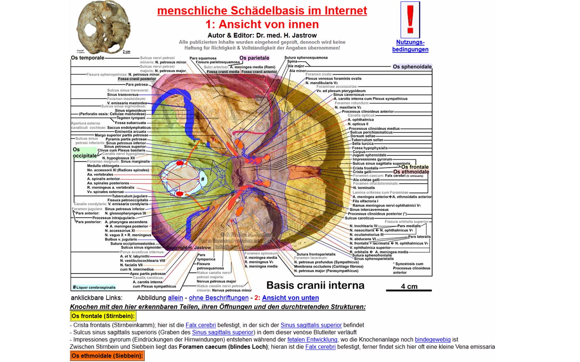 |
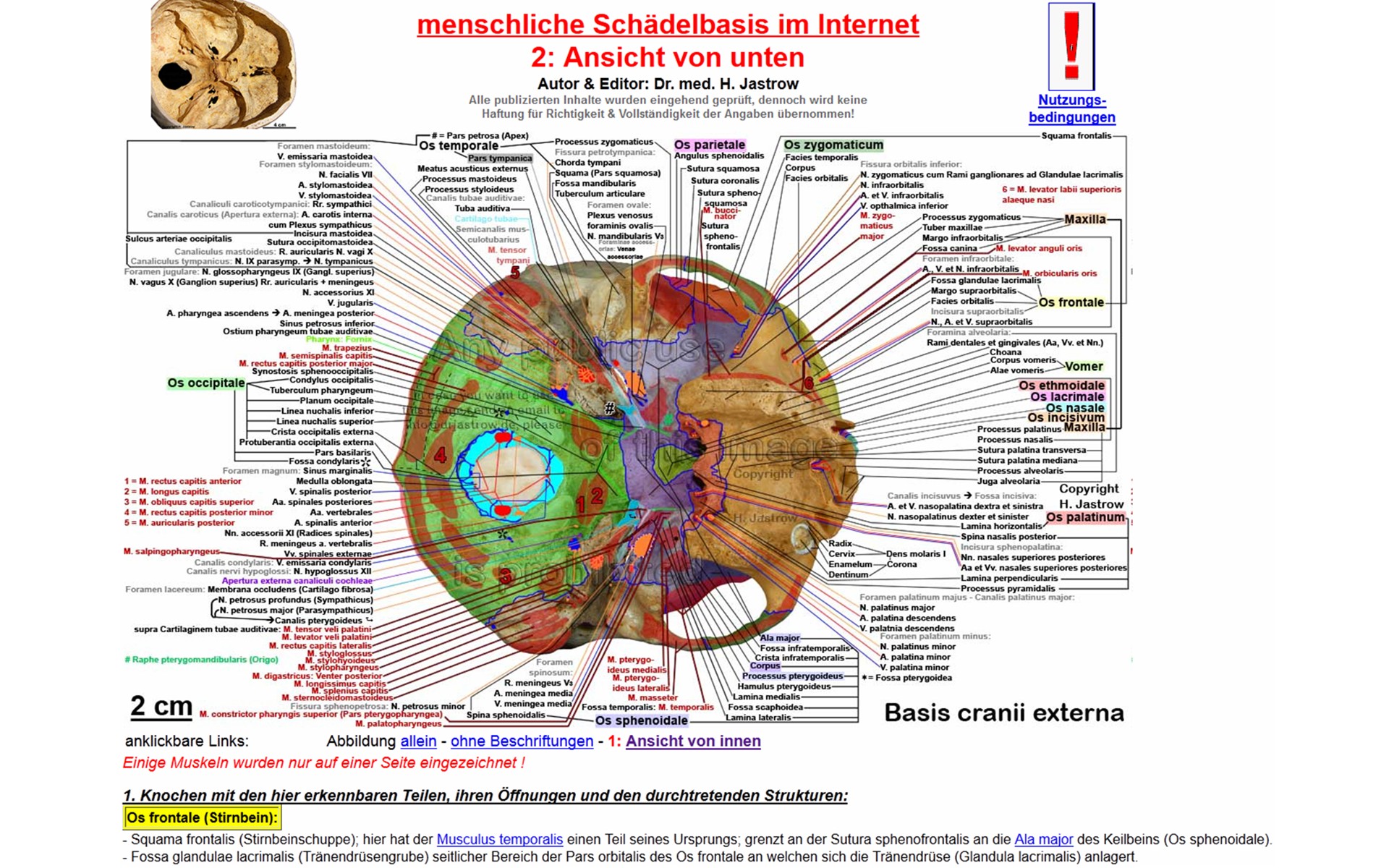
to a thoroughly labelled skull base seen from interior (Fig.37) or exterior (Fig.38) also providing detailed information on bones, their foramina and passing structures in explaining texts below. | Fig.38 - original
WAI page
 I I |
Fig.39
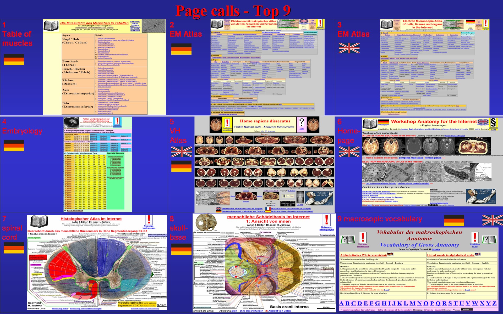 |
The top 9 pages of the internet offer from point of view of page calls
altogether are shown in Fig.39. The table of muscles and the electron
microscopic atlas is followed by human development and the cross sectional
atlas.
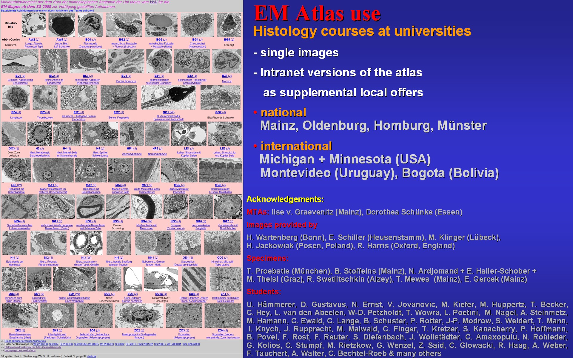
As you can see in Fig.40 high quality versions of the materials included in the internet offer are used by several universities in Germany and world-wide. On the left you see the choice of images used in the histology course of the University of Mainz, Germany. |
Fig.40 - original
WAI page
 I I |
Fig.41 - original WAI pages
1
- 2 - 3
 |
By the points listed in Fig.41 the workshop intends to be a
most useful reference for medical education and research. Besides a comprehensive
electron microscopic atlas and structured interlinked vocabularies it enables
a visual journey through the whole body from head to feet.
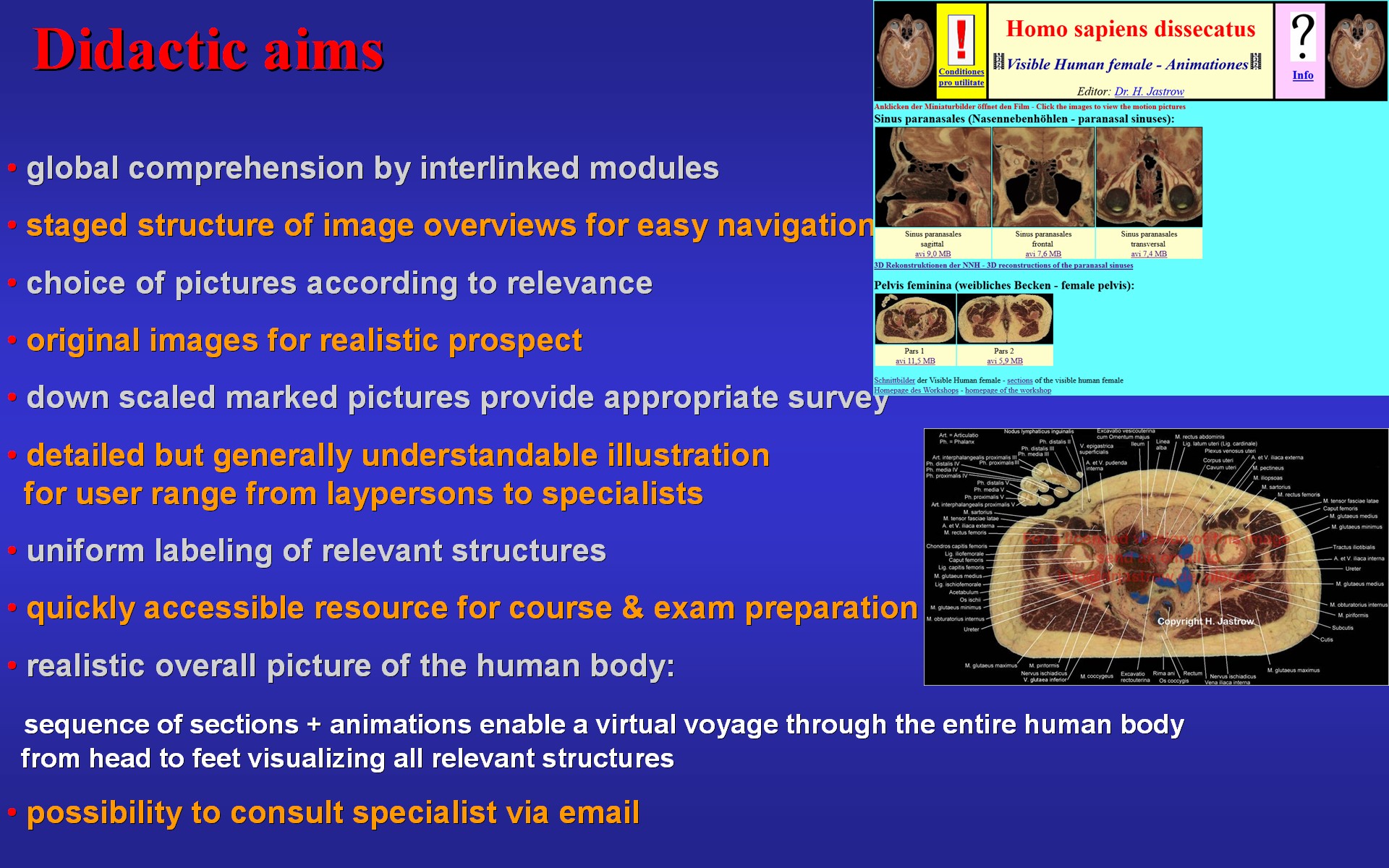
Fig.42 demonstrates the didactic aims of the anatomy offer: original images shall give a realistic aspect of true anatomy and scaled down uniformly labelled pictures shall as well as the texts provide crucial information to a user spectrum ranging from laypersons to specialists. |
Fig.42 - original WAI
pages 1 - 2
 I I |
Fig.43 - original WAI pages
1
- 2 - 3
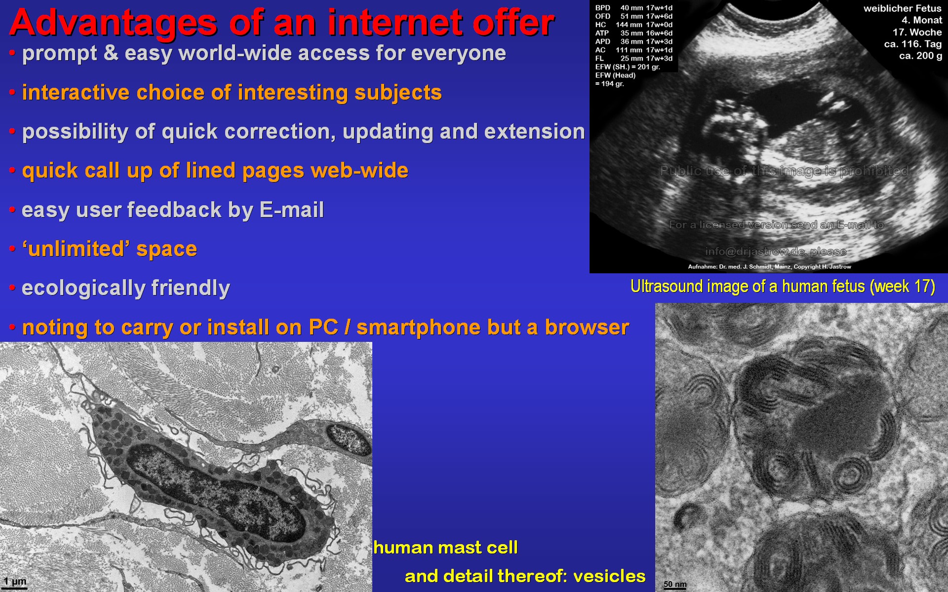 |
Fig.43 points out some of the advantages of an internet offer
like world wide availability even on a small smartphone, easy and quick
possibility to correct, to link or extend.
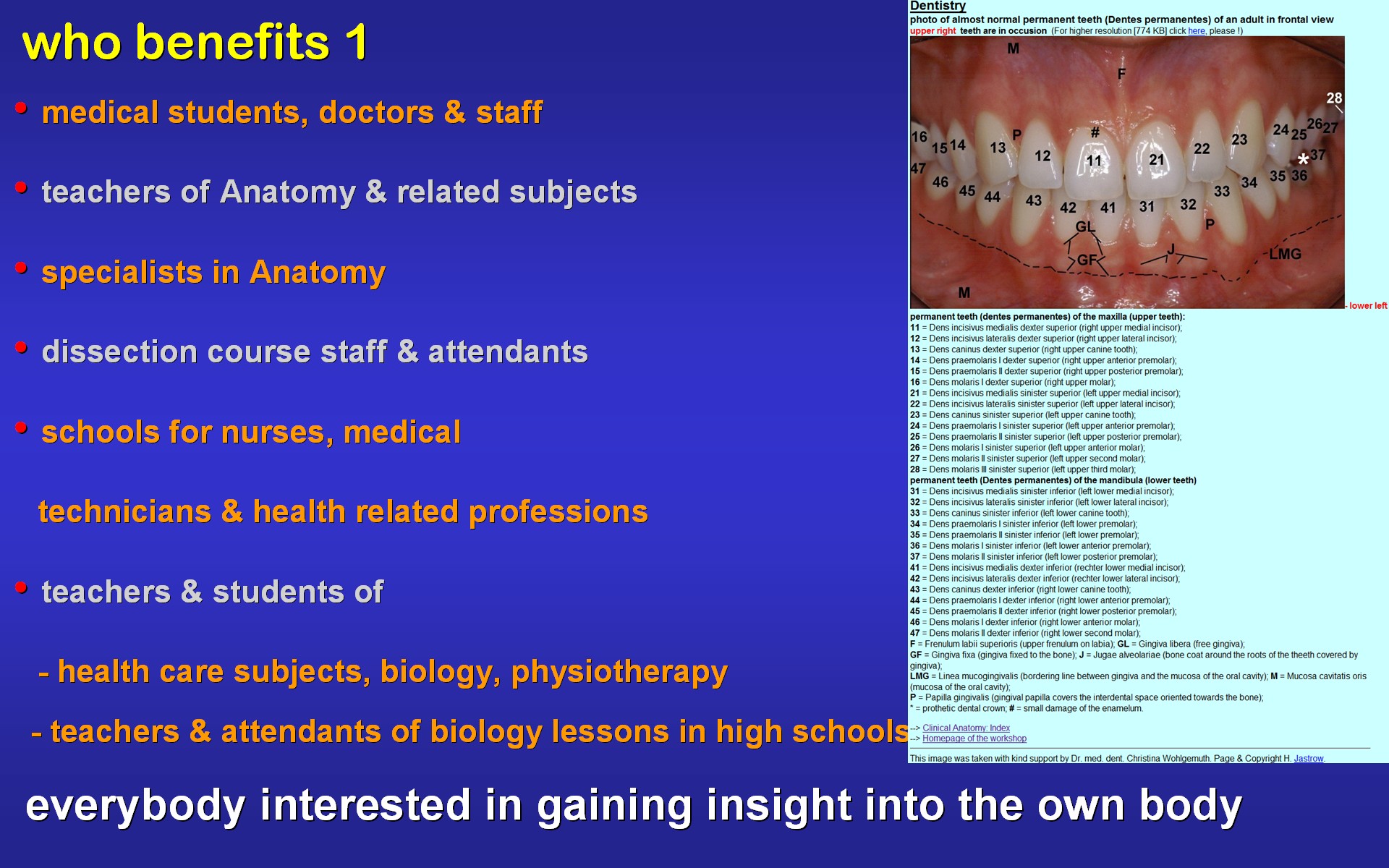
As Fig.44 constates many people can benefit from the internet atlas since the offered material can be used for teaching as well as learning anatomy in general. The spectrum reaches from universites, health care related schools and colleges to lay people interested in the construction of the human body. |
Fig.44 - original
WAI page
 I I |
Fig.45
 |
The students who helped to create some of the teaching material for
others by labelling the images in the workshop benefit in many ways and
acquire a special qualification useful for their later work as medical
doctors and when seeking positions (Fig.45).
The offered material is suitable for study and instruction of anatomy in general. In Fig.46 you see some examples what it can be used for. It enables self-assessment of knowledge and may even serve as reference for scientific purposes. The clinical images help pupils and patients to understand anatomy. The quick overviews on different topics and the special interest pages are frequently used for exam preparation. |
Fig.46 - original WAI
pages 1 - 2
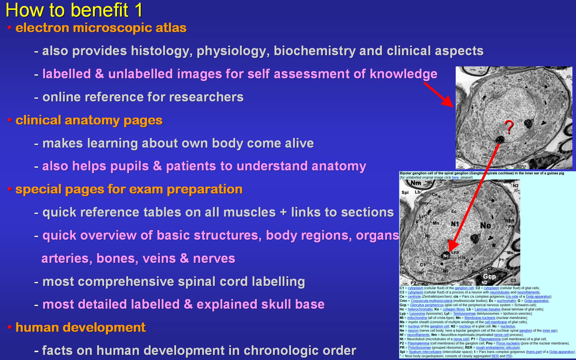 I I |
Fig.47 - original
WAI page
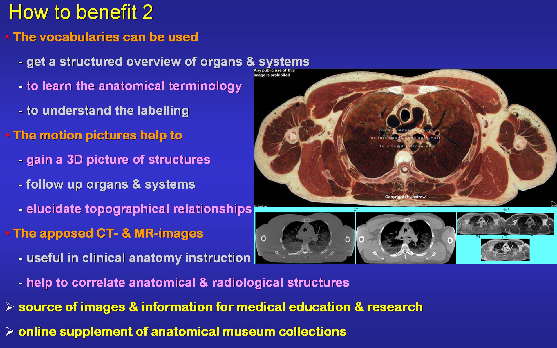 |
For non-professionals the vocabularies are valuable to understand the
terminology. The sequence of sections and motion pictures facilitate acquisition
of a 3D-understanding of anatomy and topographical relationships. A favoured
feature of the cross-sectional atlas is the easiness of correlation of
original section to corresponding radiological images. In brief the atlas
is a source for many uses (Fig.47).
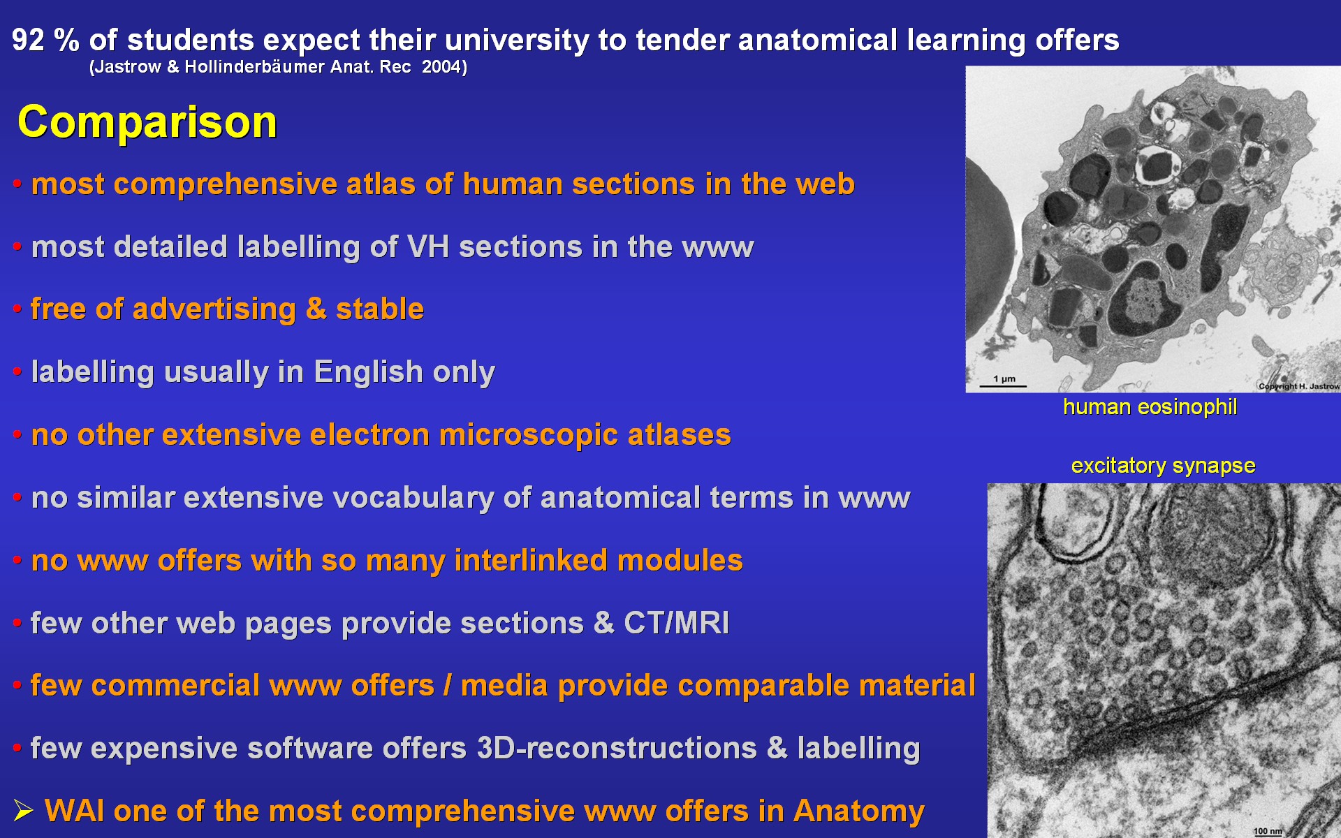
Today students demand good teaching offers in this context the present atlas the most comprehensive one in the internet offering most detailed labelling of sections in the web. It is free of advertising and runs on all kind of browsers. Comparable extensive electron microscopic material is only available in few commercial textbooks. With its further anatomy teaching offers the workshop Anatomy for the internet represents a unique concept of presenting anatomy to specialists as well as to the general public (Fig.48). |
Fig.48 - original WAI pages
1
- 2
 I I |
Fig.49
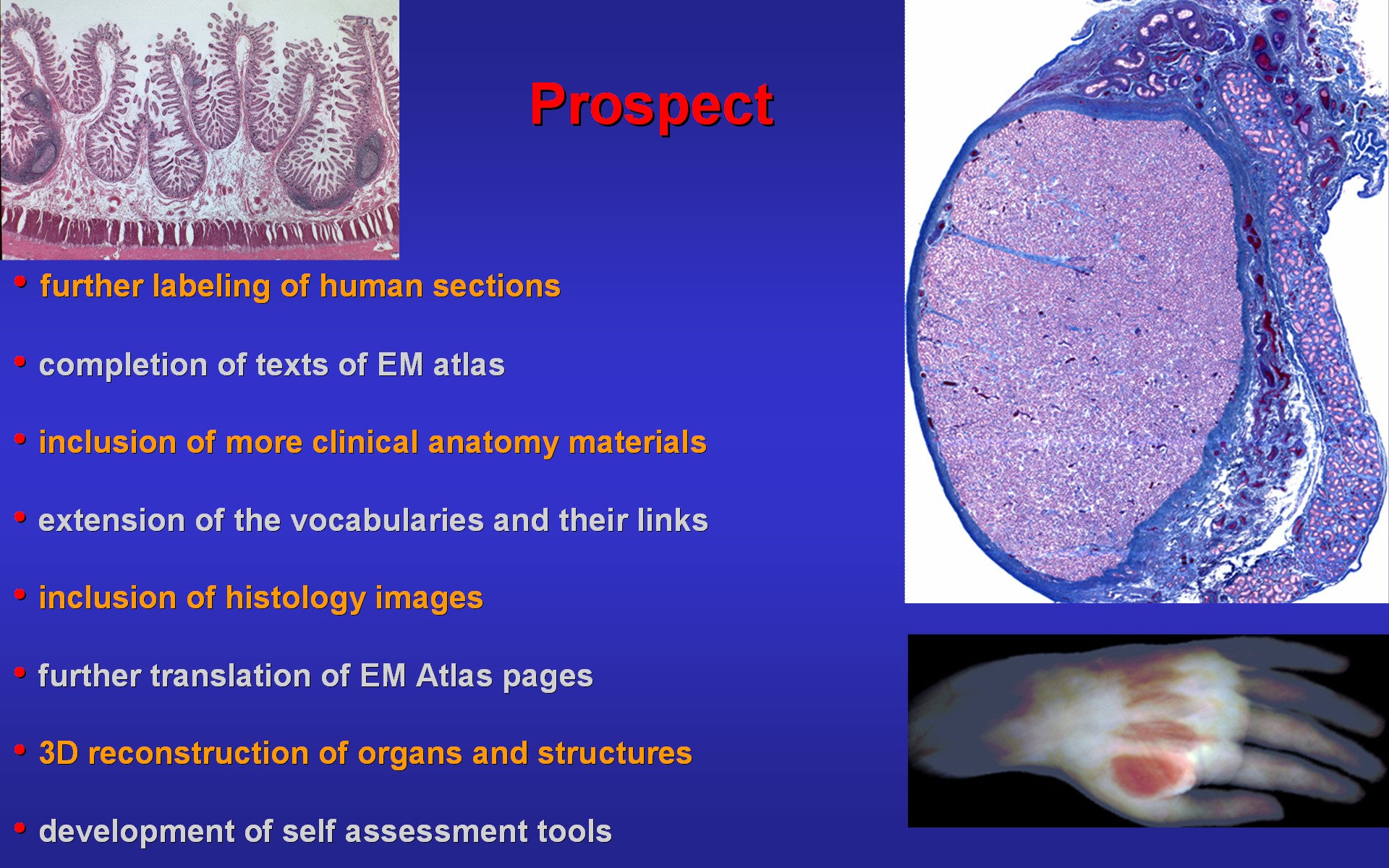 |
The present offer is constantly extended by including further materials.
Presently the texts of the EM Atlas are completed and first histology pictures
are integrated. Further translation of still German pages is going on.
On a longer time scale 3D reconstructions and self assessment tools are
planned. With these goals it is intended to contribute to world-wide improvement
of understanding of anatomy in pre-clinical and clinical medicine (Fig.49).
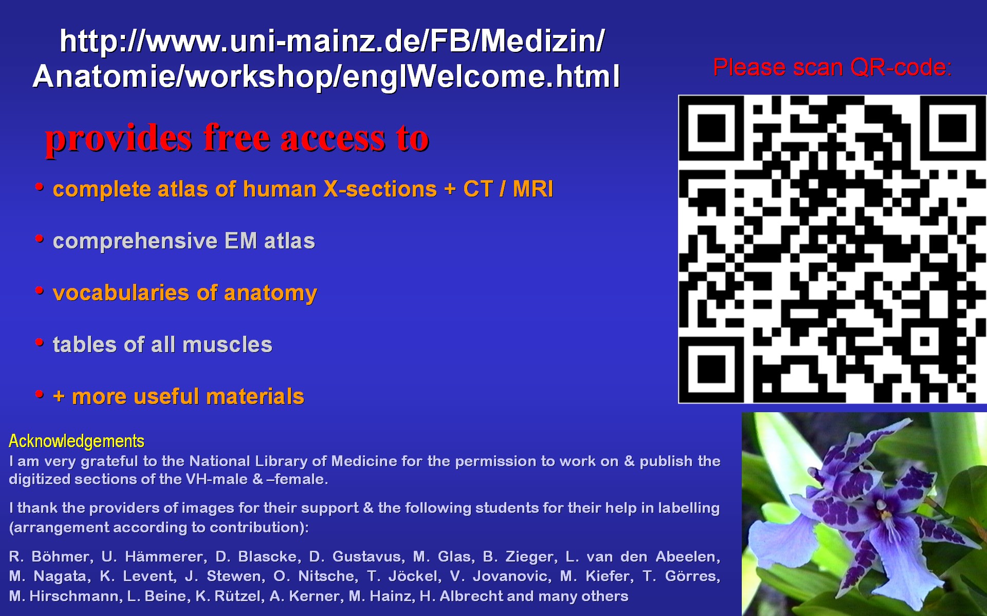
With the short summary in Fig.50 I kindly to invite you to explore the internet pages of the workshop which you can easily access when scanning the shown QR-code. Further, I acknowledge those who supported this project. |
Fig.50 - linked
WWW page
 I I |
Fig.51 - linked
WWW page
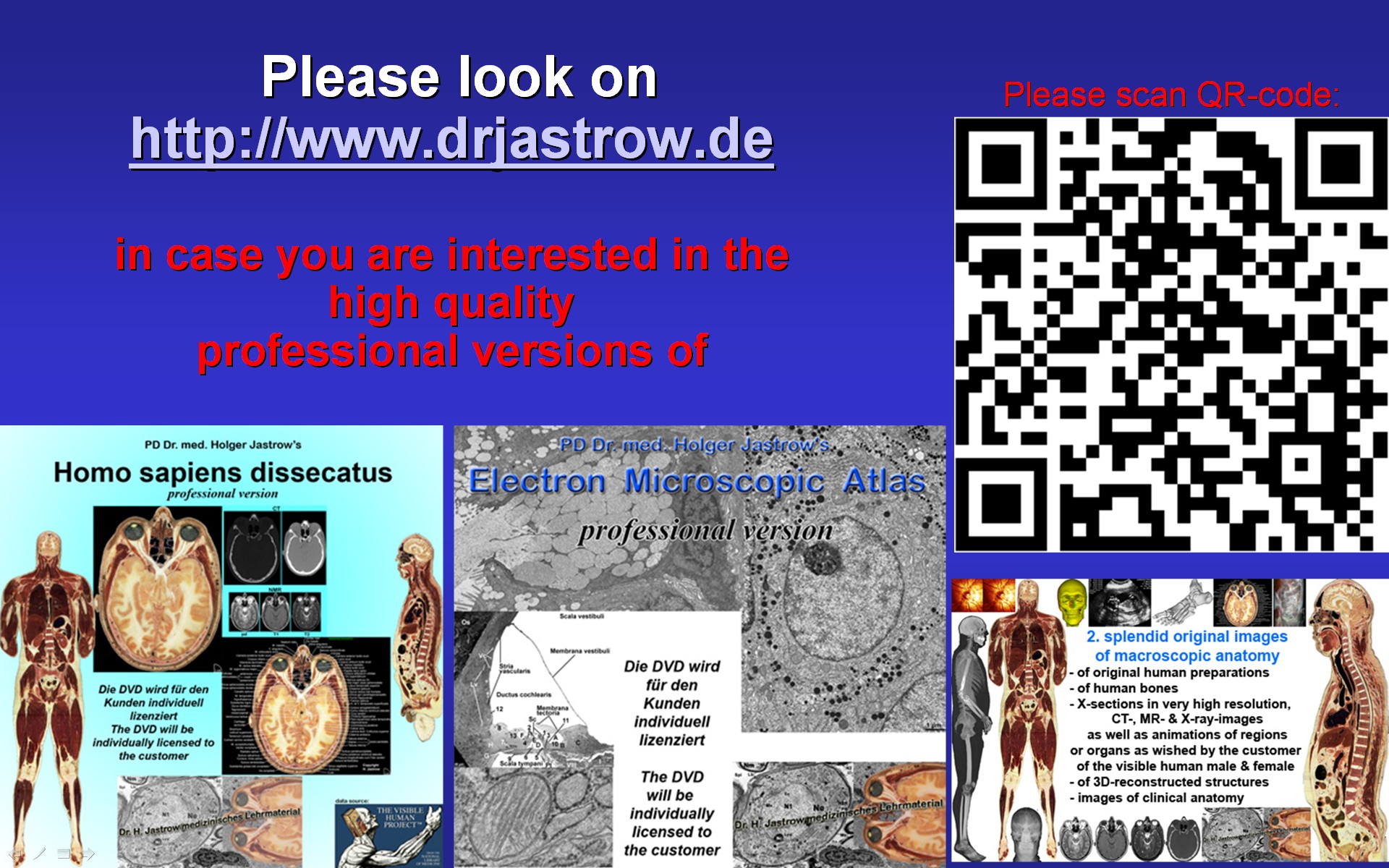 |
Please note that there are also high quality professional versions
of both atlases available (Fig.51).
Thank you for your attention! |
In case you are interested in any
material of the WAI,
please contact specialist in Anatomy Kotthaushang 22, 45239 Essen, Germany Email: info@drjastrow.de; Internet: http://www.drjastrow.de Phome: +49 201 4749893 |