List of abbreviations
Vocabulary
of micros-
copic
anatomy
specialist terms
explained in
English +
German

Every attempt was made to provide correct information and labelling, however any liability for eventual errors or incompleteness is rejected!

dieser Seite

Editor:
Dr. med.
H. Jastrow


Conditions
of use
Overview human retina (Retina):
Pages with explanations are linked to the text below the images if available! (Labelling is in German)
The retina of the eye (Terminologia histologica: Retina) consists of a sensory part for vision (Pars optica) and a small marginal blind zone (Pars caeca, which has a part that covers the iris; Pars iridica and a further part covering the ciliar process; Pars ciliaris). The small blind zone has a monolayered prismatic epithelium. In contrast to that the Pars optica has a very complex structure and two areas that differ from the general composition descibed below: the papilla of the optic nerve (Discus / Papilla nervi optici = blind spot; diameter ~ 1.7 mm) and the yellow spot (Macula lutea).
The optic neve leaves the eye in the region of the Papilla nervi optici, further the retinal vessels (Arteria and Vena centralis retinae) enter the ocular bulb here. Due to this no sensory cells but only non-myelinated nerve fibres and vessels are present in this region (see clinical image).
Lateral (temporal) thereof the Macula lutea, which is ~3 mm in diameter, is located in the centre of the retina. It has a central funnel-like depression, the central fovea (Fovea centralis; diameter ~ 1.5 mm), which is the spot with maximal visual acuty, i.e. the highest spatial resolution. The Foveola is located in the centre of the fovea. It is the thinnest area of the pars optica, has a diameter of 0.35 mm and only consists of cones and processes of Müller glial cells. Incoming light is not dispersed by covering layers in this area. The foveal cones have a 1:1 connection to ganglion cells this allows highest spatial. The number of rods which first appear in the foveal area increases towards the periphery; in surrounding macula rod and cone number is about equal then rods rapidly increase while cones decrease.
When regarding the Pars optica of the retina from outside to inside, the following layers can be distinguished:
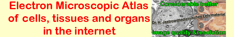
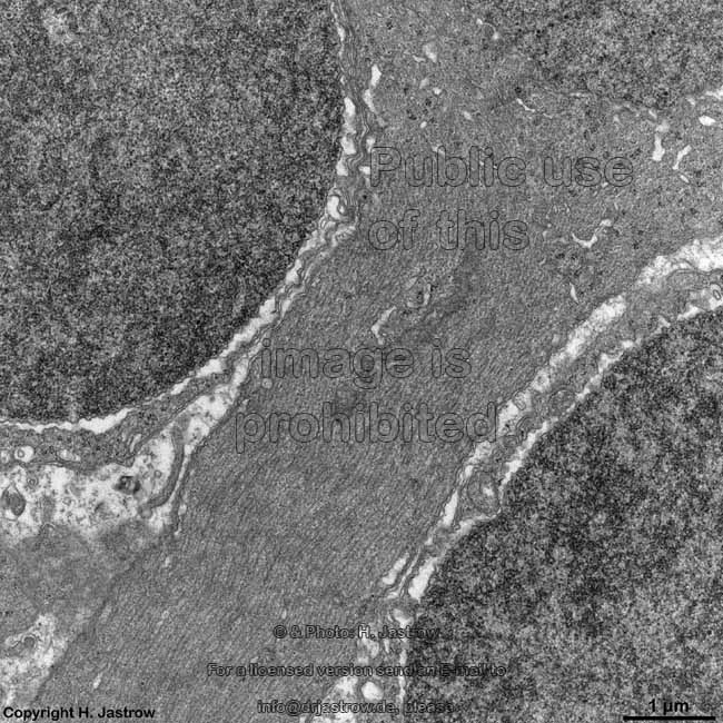
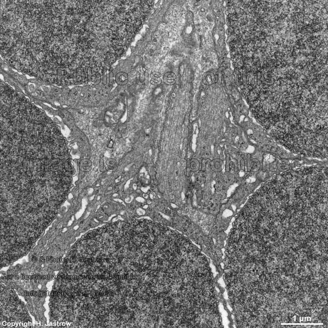
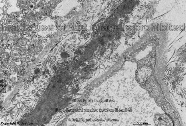
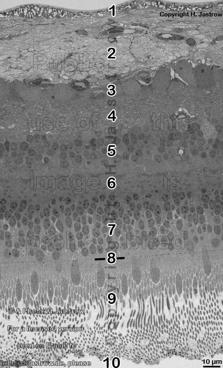
1. inner limiting membrane = Stratum limitans internum (--> images) borders the vitread body (Corpus vitreum) with a basal lamina which has considerable regional differences in thickness, radial fibres spread above the basal lamina which are the endings ofMüller Glial cells. They are connected to each other via small tight 
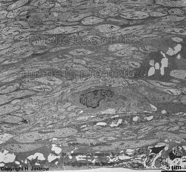
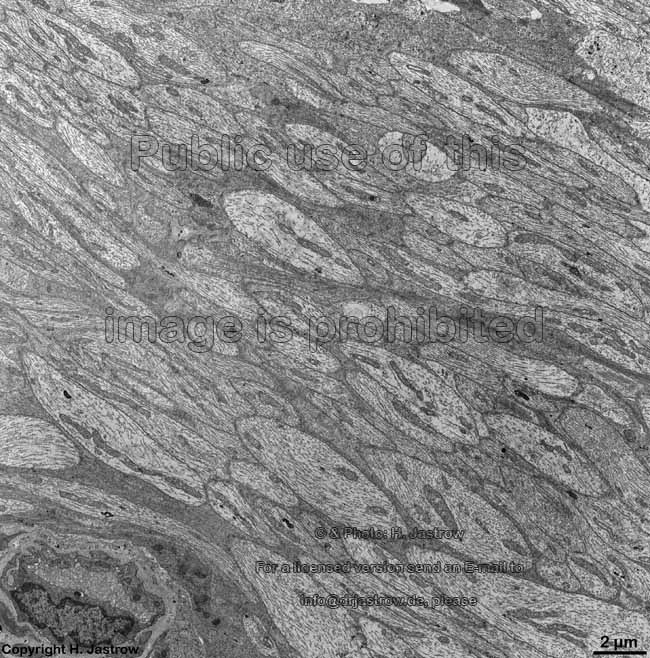
junctions just above the basal lamina.
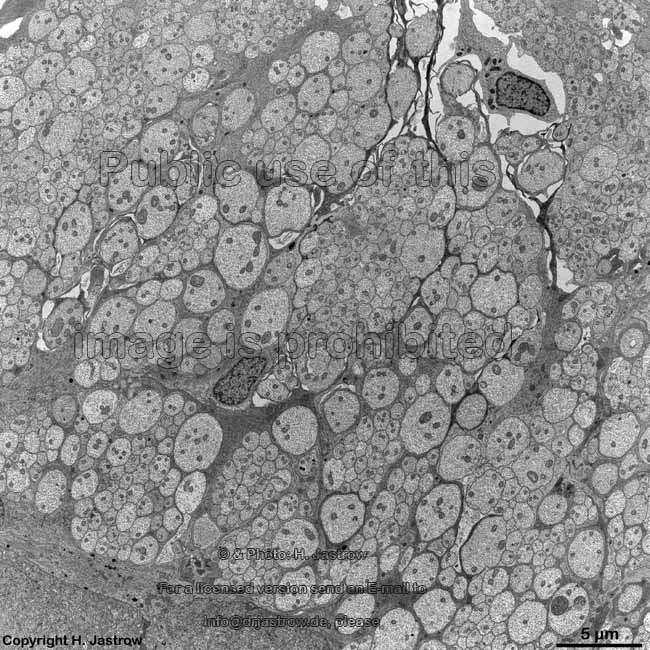
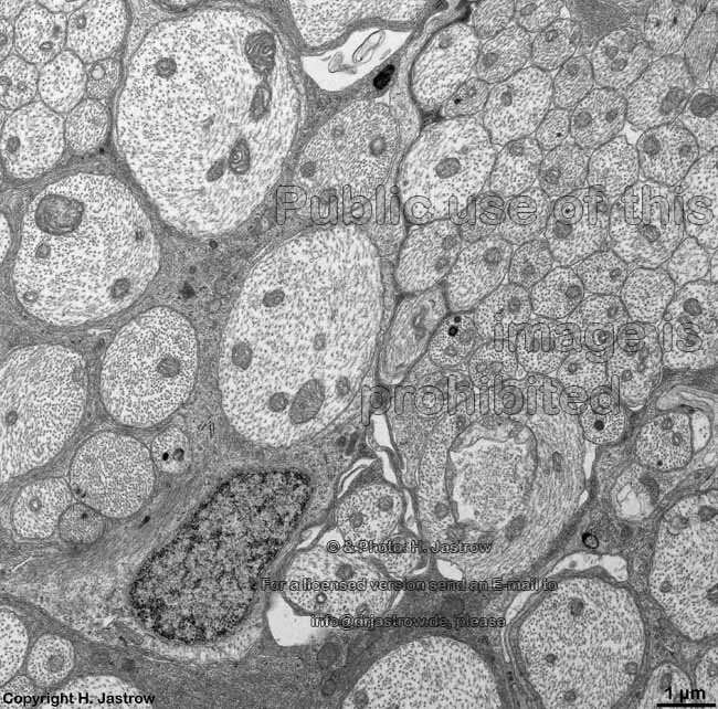
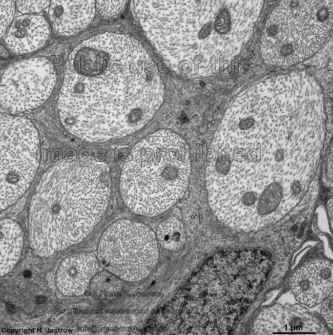
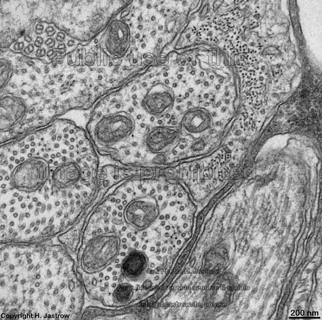
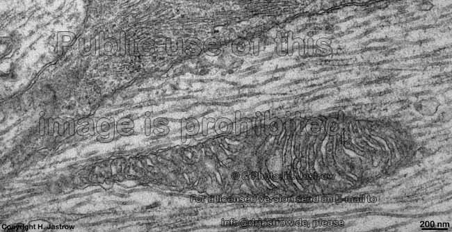
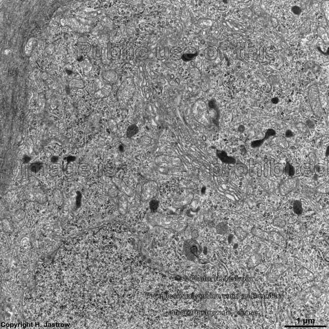
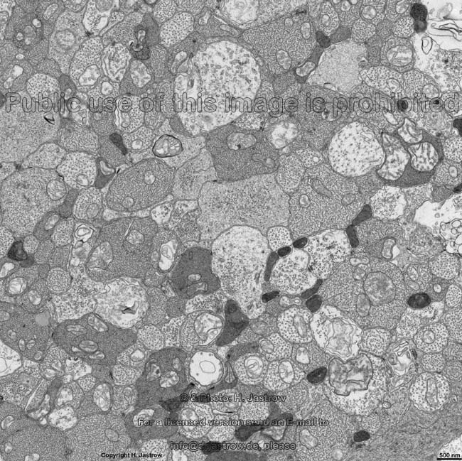
2. nerve fibre layer = Stratum neurofibrarum (--> images) virtually all non-myelinated axons which form the optic nerve at the papilla, further some blood vessels are present here
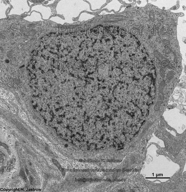
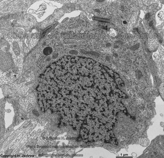
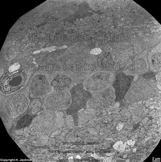
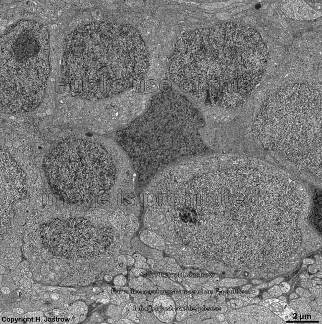
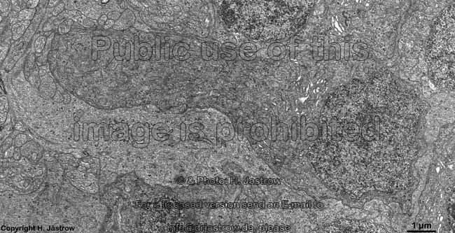
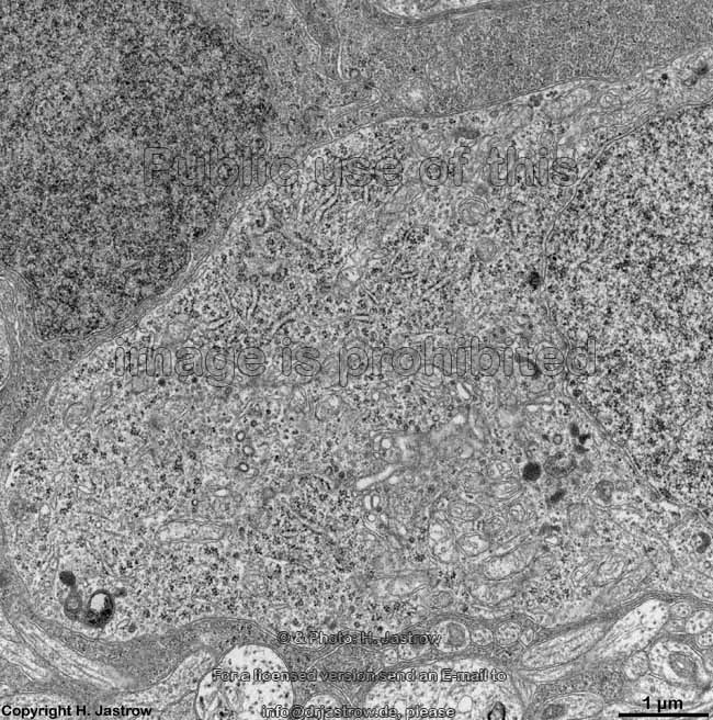
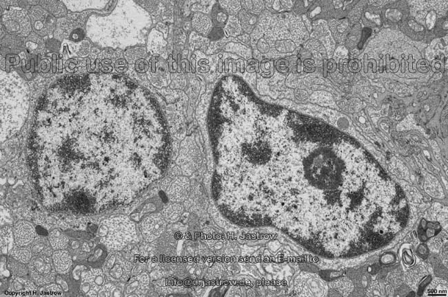
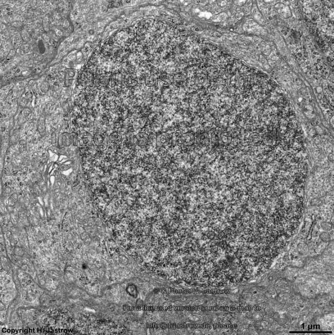
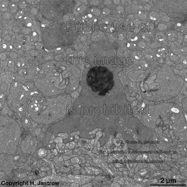
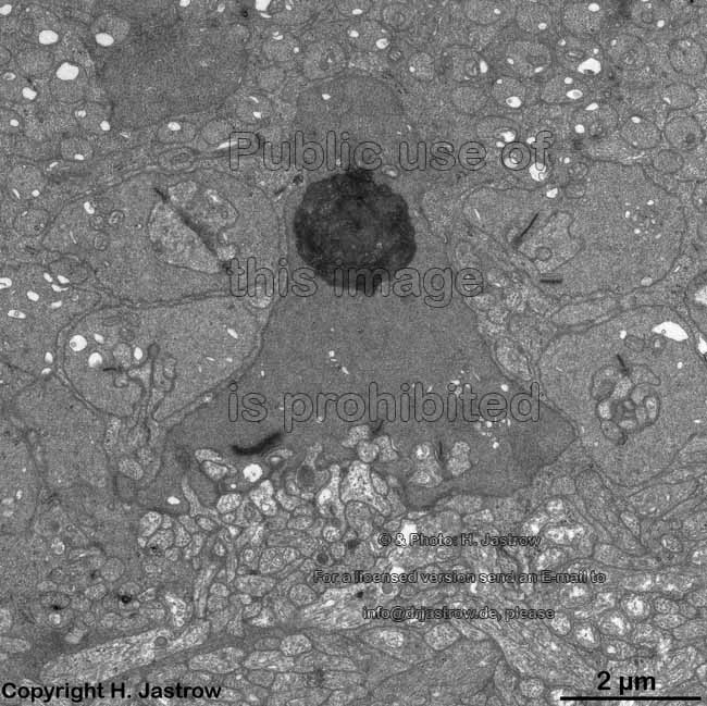
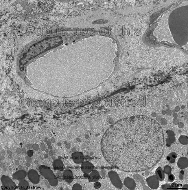
3. ganglion cell layer = Stratum ganglionicum (--> images) with multipolar ganglion cells, which are the third neurons of the visual pathway
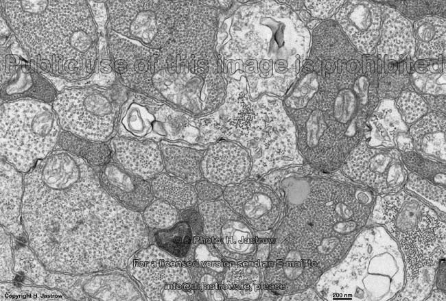
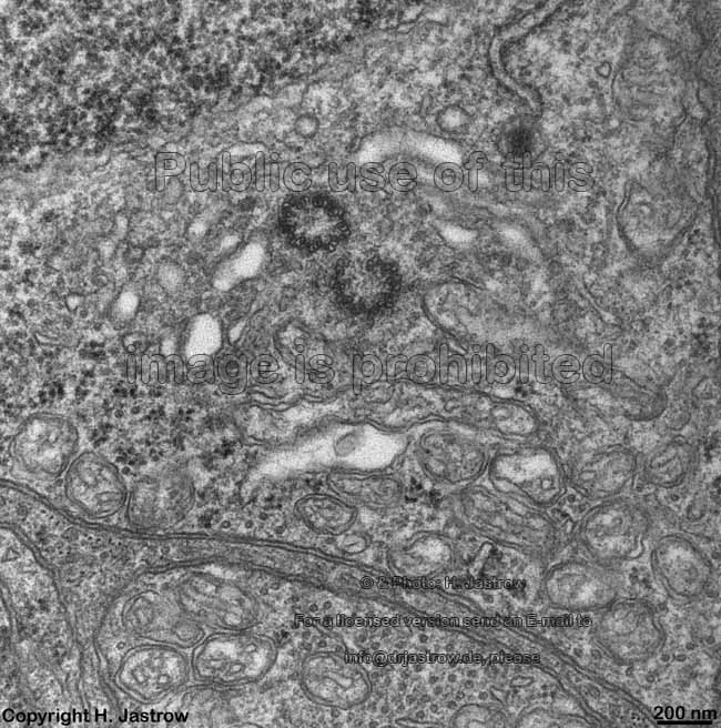
4. inner plexiform layer 
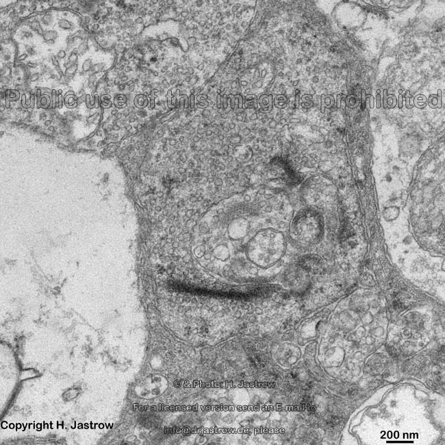
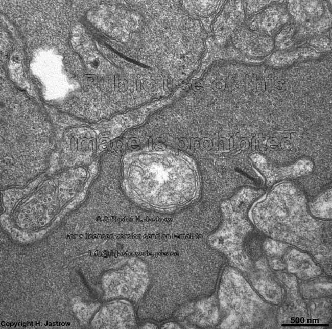
= Stratum plexiforme internum (--> images) contains the synapses between the second (bipolar cells) and the third neurons (ganglion cells) of the visual pathway. Most of these synapses are ribbon synapses with synaptic bodies, further many conventional chemical and a few electrical synapses are present here.
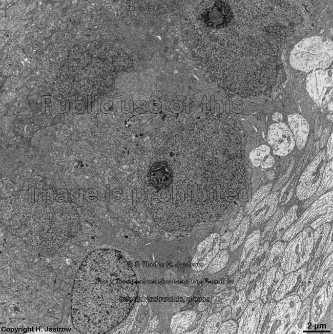
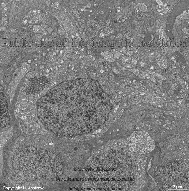
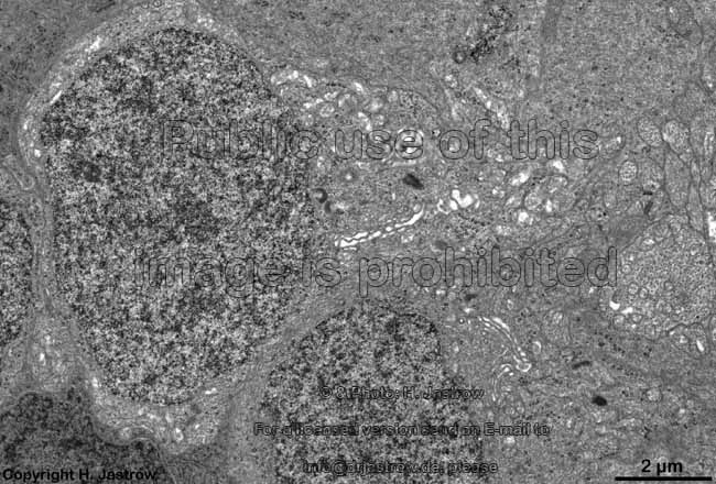
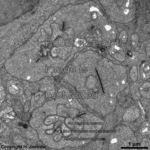
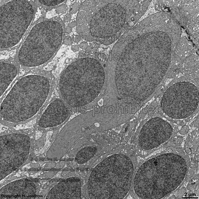
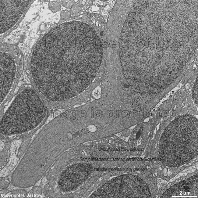
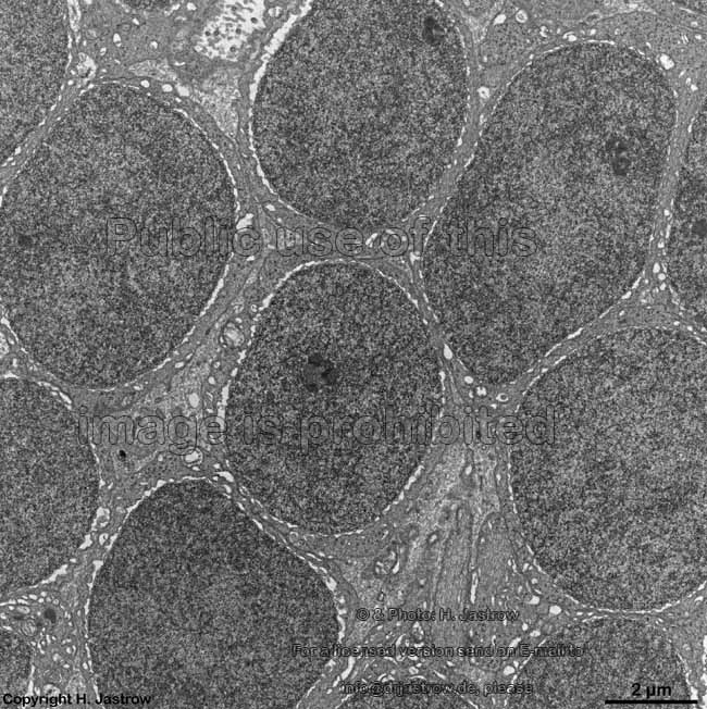
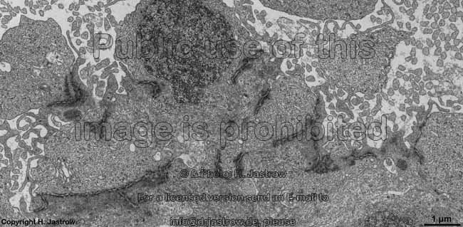
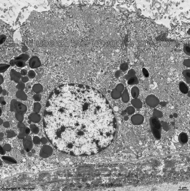
5. inner nuclear layer = Stratum nucleare internum (--> images) with perikarya of A. bipolar cells (rod- and cone bipolar cells = second neurons of the visual pathway which can be further callsified in many fuctionally different types), B. some amacrine cells, which are located at the border to 4, C. horizontal cells, which are also less frequent and located at the border to 6. Some of the horizontal cells show very large aggregated macrotubules in their cytoplasm. D. some intermingeled nuclei of radial fibre cells, i.e. Müller's glial cells.
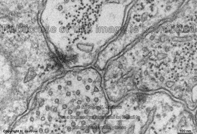
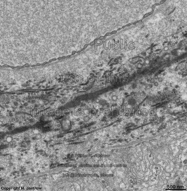
6. outer  plexiform layer = Stratum plexiforme externum (--> images) 
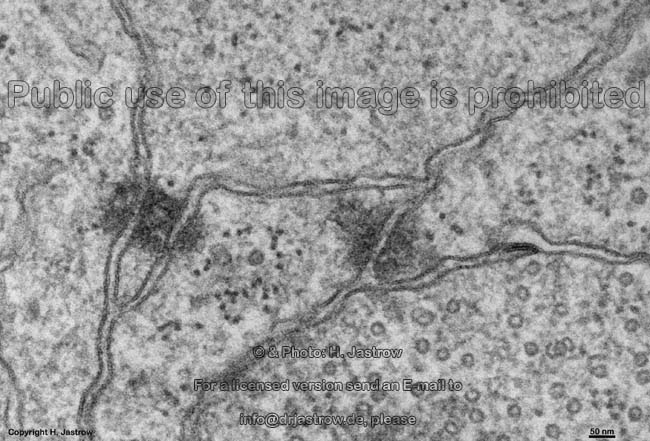
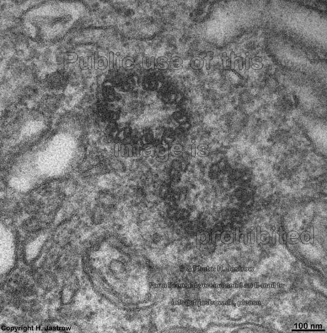
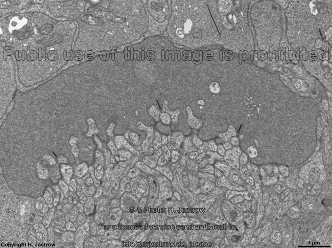
Processes of horizontal and bipolar cells are invaginated into the terminals of the photoreceptor cells (rods and cones) that show special electron-dense cell organelles, the synaptic ribbons, close to their presynaptic membranes in this area. The resulting ribbon synapses serve for ultrafast (tonical) signal transduction to the dendrites of the second neurons of the visual pathway (rod- or cone bipolar cells).  Two bizarrely formed horizontal cell processes with terminal swellings are present in one invagination of rod terminals that often is subdivided in two endings. Whereas these horizontal cell terminals are located laterally, 1 - 3 thin bipolar cell processes comprise the centre of a ribbon synapse. In contrast to that cone terminals always show 25 up to over 50 invaginations with usually two lateral horizontal cell processes around 1 - 2 short bipolar cell dendrites. Apart from the majority of ribbon synapses, conventional chemical synapses are present outside the invaginations. Electrical synapses (gap junctions) are rare in this layer.
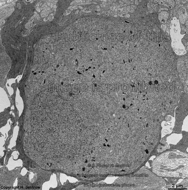
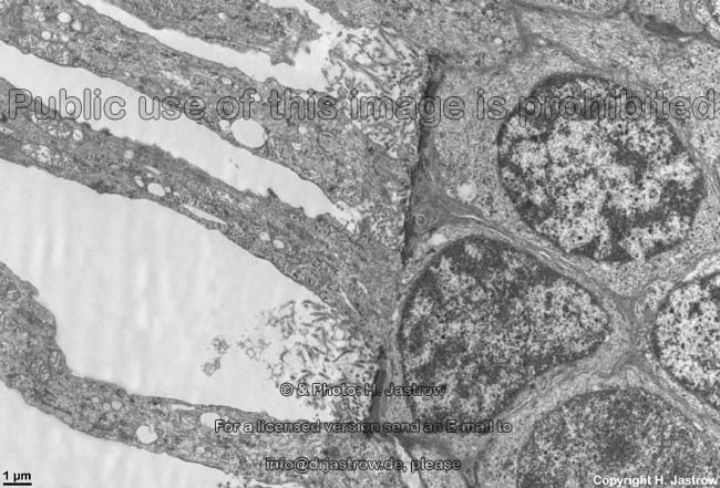
7. outer nuclear layer = Stratum nucleare externum (--> images) with the nuclei of the photoreceptor cells (rods and cones = first neurons of the visual pathway)
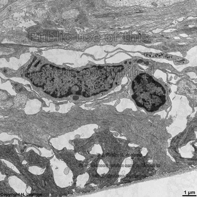
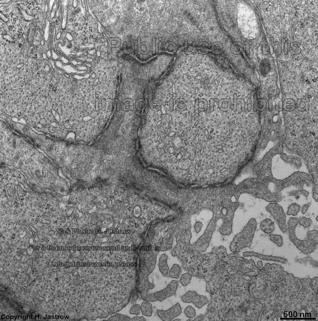
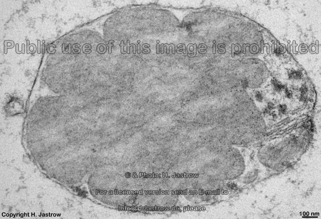
8. outer limiting layer = Stratum limitans externum (--> images) area with special belt desmosomes (Zonulae adhaerentes) located between the receptor cells and the very narrow terminals of Müller's glial cells
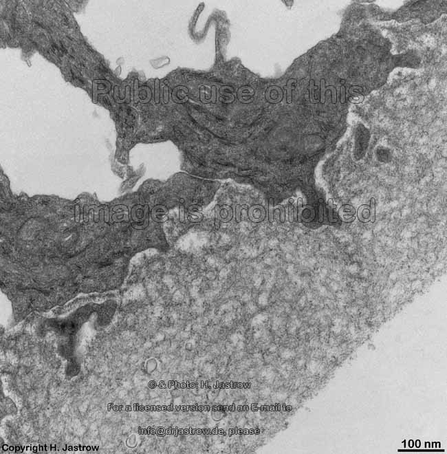
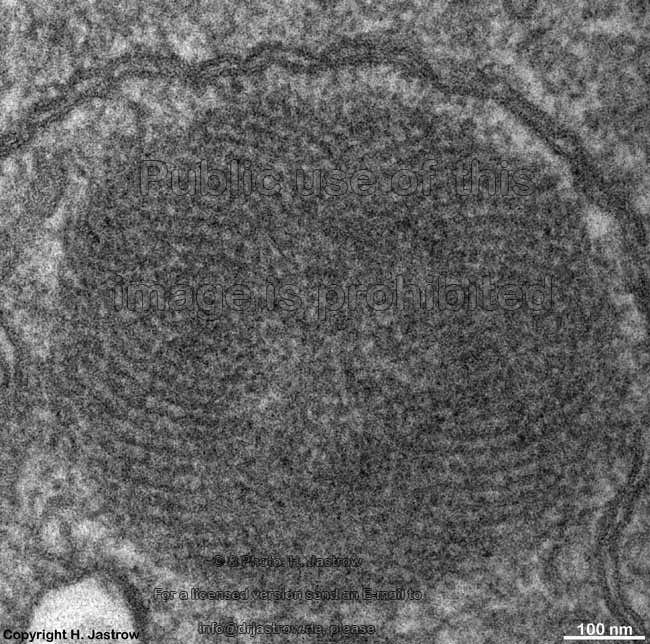
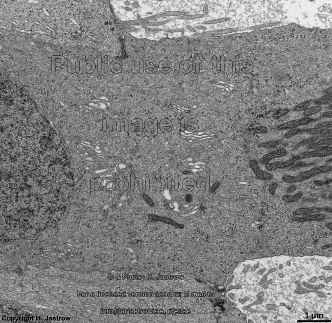
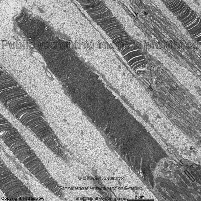
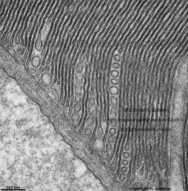
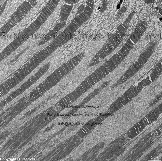
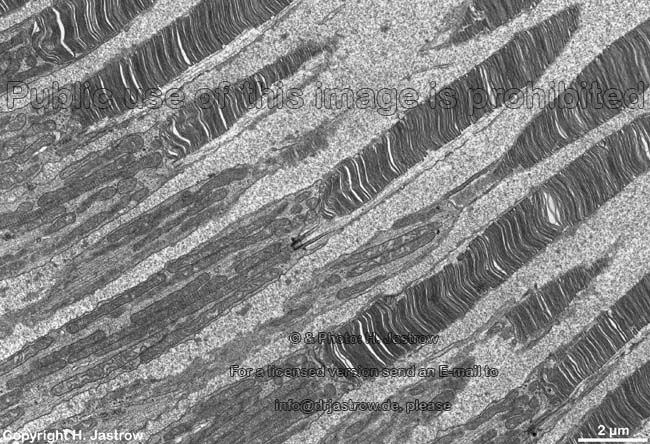
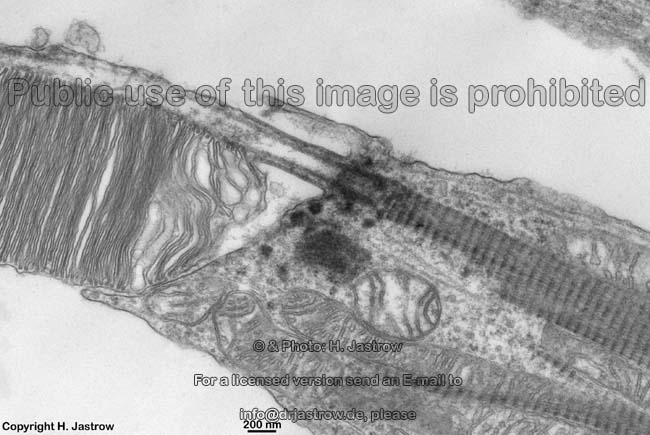
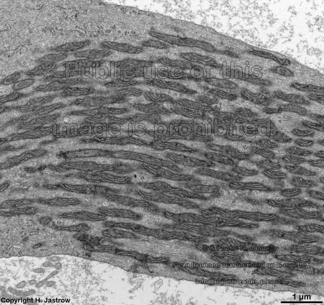
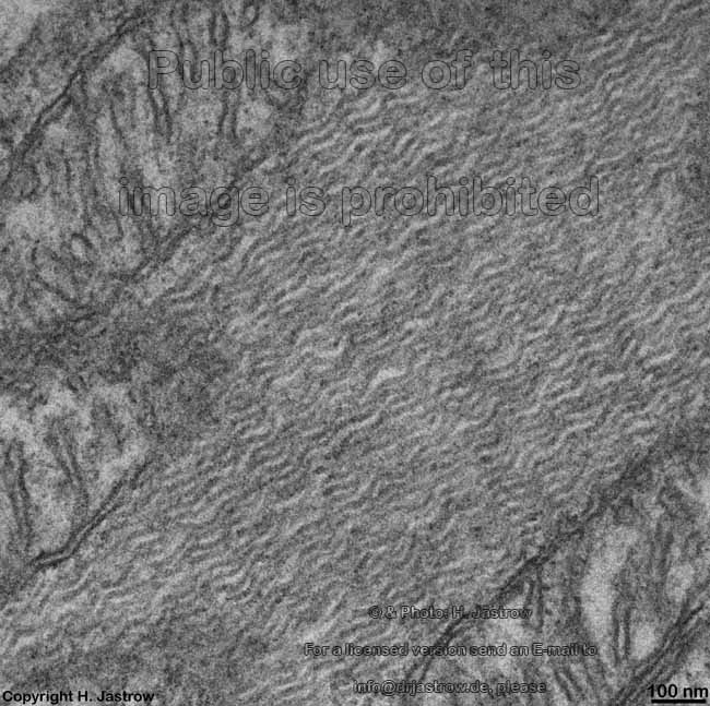
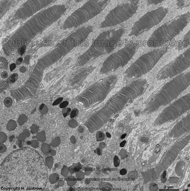
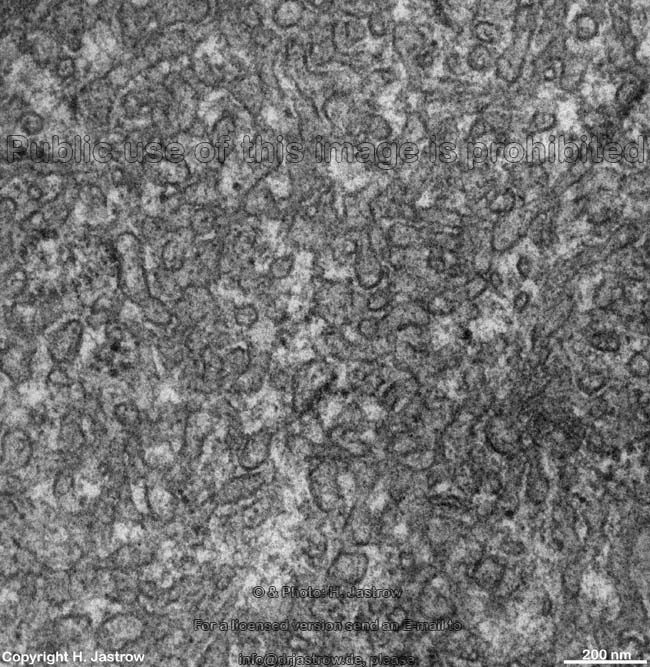
9. layer of inner and outer segments of rods and cones = Stratum segmentorum externorum et internorum (--> images) upper part: inner- and lower part: outer segments of rods and cones.
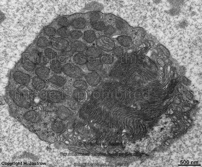
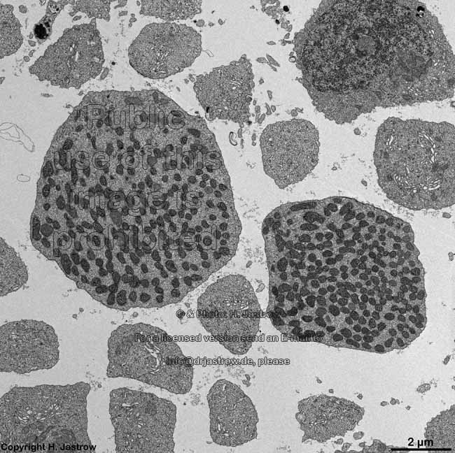
The Ellipsoid is the part of the inner segment of a photoreceptor that is closer to the outer segment. It is very rich in mitochondria, shows some root fibres, wave-like bundles of intermediate filaments and microtubules. 
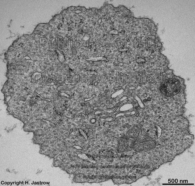
The ellipsoid continues into the Myoid, the lower part of an inner segment which is rich in Golgi-apparatuses and RER but hardly shows any mitochondria. 
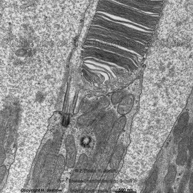
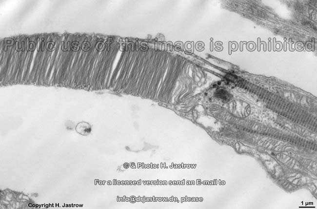
The inner and outer segments are surrounded by a liquor filled space into which long, thin processes of Müller's glial cells reach from outside whereas from inside other long, thin processes of pigment epithelial cells protrude. The outer and inner segments are connected to each other only via a narrow cytoplasmatic bridge containing a cilium with 9x2 + 0 microtubules.
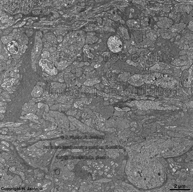
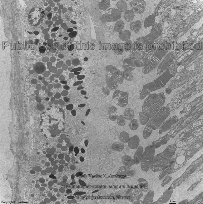
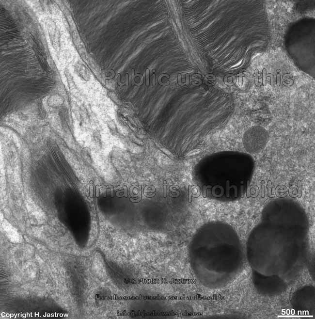
10. pigment epithelium (Stratum pigmenti = Pars pigmentosa; --> images) with pigment epithelial cells which phagocyte the tips of the rod or cone outer segments. The incorporated parts of the outer segments condense further and finally may no longer be distinguished from nearby pigment vesicles which prevent reflection of photons from incomeing light. Vast amounts of smooth endoplasmic reticulum are characteristic for the cuboid pigment epithelial cells. The junctional complexes between these cells are rich in tight junctions and the morphological base for the blood-retina barrier.
Underlying Bruch's membrane, a rather thick basement membrane with lots of elastic and collagen fibres comprises the border to the Lamina choroidocapillaris (--> images) which has lots of blood vessels. The subsequent choroidea shows pigment epithelial cells scattered in loose connective tissue with an abundance of smaller blood vessels (especially fenestrated capillaries and venoles). The outermost layer of the ocular bulb is the strong sclera which consists of thousands of very tight parallel ordered collagen fibre bundles.
functional aspects:
The human retina has about 6 000 000 cones and 120 000 000 rods, whereby the concentration of cones diminuishes considerably from the foveola to outside. The very long outer segments of the photoreceptor cells show highly ordered membrane discs which reach forward into the pigment epithelium. The visual pigment. which is able to percept photons of the visible light, is located in these discs but too small to be visible in the electron microscope. It is rhodopsin in rods which consists of the glycoprotein opsin and the bound pigment 11 cis-retinal as chromophor. Rhodopsin is a transmembrane proteine, i.e. directly incorporated in the disc membrane. It is synthetised from vitamin A (retinol). Formation of the membrane discs is continious and it takes 10 days to renew a complete outer segment. Formation starts at the border to the inner segment. The small cytoplasma bridge between inner and outer segment is stabilised by a simple cilium (no kinocilium, only 9x2 outer microtubules but no inner pair of microtubules; see images). Rods are responsible for perception of small amounts of light and darkness (viewing of grays at dusk = scotopic vision with inferior spatial resolution). Humans have 3 different kinds of cones for perception of different colours. Cones react to colour and colour intensity and have more complex visual pigments. Due to a very complicated interconnection of different cell types the spatial resolution of cones is much better than that of rods. However this photopic vision requires more light (intensity) than scotopic vision. The pigments involved in colour vision of cones have different absorption maxima of wave lengths of light which are the basis for all the about 7 million different colours humans may percieve. The wave lengths for the basic colours are: 700nm IIII Red, 546 nm IIII Green, 435 nmIIII Blue. All other colours are generated by different intensities i.e. mixtures of the basic colours. The morphological base for this is the sophisticated interconnection of the different visual cells. Some examples: yellow is generated by red and green in same intensity; white = red + green + blue in maximal intensity, otherwise grays are generated, black = no wave lengths. All colours may be percieved at different intensities (as it is the case for RGB values of monitors). With the exception of the central region (fovea) one bipolar cell always sends dendrites to several receptor terminals and several bipolar cell axons reach one and the same multipolar ganglion cell. The axons of these ganglion cells collect to fibre bundles which form the optic nerve (Nervus opticus = brain nerve II) which runs to the brain.
 
inner limiting membrane = Stratum limitans internum (1)
Overview with a part
of the nerve fibre layer
Müller cell processes with
wide intercellular spaces
2 Müller cells in
a basal location
inner limiting mem-
brane proper
detail: thick
basal lamina
nerve fibre layer = Stratum neurofibrarum (2)
Müller cell process
and nerve fibres
nerve fibre layer
overview (X-section)
detail thereof with 
Müller glia cell
 further detail
thereof
non-myelinated axons
of ganglion cells
nerve fibre layer overview
in longitudinal section
nerve fibre layer in an
oblique overview
detail: dark mitochondrium of the crista-
type in the axon of a ganglion cell
Müller's glia cell 1 Müller's glia cell 2
Ganglion cell layer = Stratum ganglionicum (3)
ganglion cells 
overview
ganglion cell 1 cytoplasm thereof ganglion cell 2 Müller cell + 
ganglion cells
inner plexiform layer = Stratum plexiforme internum (4)
inner plexiform 
layer 1
detail of the inner 
plexiform layer
inner plexiform 
layer 2
Puncta adhaerentes of the 
inner plexiform layer 1
Puncta adhaerentes of the 
inner plexiform layer 2
synaptic bodies (ribbons and spheres) in
axons of bipolar cells (animation)
inner nuclear layer = Stratum nucleare internum (5)
Overview horizontal cell with 
Macrotubuli aggregati
nuclei of bipolar 
and Müller cells
nuclei of
different cells 1
nuclei of
different cells 2
rod bipolar cell with an ectopic synaptic 
ribbon in a dendritic process
cone bipolar cell with large dendrite
nuclei of
different cells 3
amacrine cell 1 amacrine cell  2 nucleus of an
amacrine cell 
nuclei of 2 bipolar cells centriols of a
bipolar cell
detail thereof bipolar cell nucleus
outer plexiform layer = Stratum plexiforme externum (6) with synaptic ribbons
rod terminals, outer 
plexiform layer
crystal close to a
ribbon synapse
detail: crystal rod terminal 
(4 h post mortem)
cone & rod terminals 
serial image 1
cone & rod terminals 
serial image 2
cone & rod terminals 
serial image 3
ribbon synapses 
rods & cone
cone terminal with multiple 
SBs, outer plexiform layer
Animations of ribbon synapses with postsynaptic elements in terminals of rods and cones which are located in this layer (click an image to start the animation !)
1 cone- & 4 rod terminals stereo animation of a rod ribbon synapse ribbon synapses and postsynaptic processes of a cone 
outer nuclear layer  = Stratum nucleare externum (7)
overview with cone
nucleus and axon 1
detail thereof:
axon hillock 1
other cone nucleus with
axon hillock 2
detail thereof:
axon hillock 2
similar detail nuclei mainly of rods detail thereof
outer limiting membrane = Stratum limitans externum (8)
oblique section of the zonulae 
adherentes of the outer limiting membrane
Stratum limitans ex- 
ternum (X-section)
outer nuclear layer, 
inner segments
detail myoid, Stratum 
limitans externum
similar detail rods: inner segments + nuclei 
(fixed 4 h post mortem)
layer of the outer segments, clia + inner segments of rods and cones = Stratum segmentorum externorum et internorum (9)
inner + outer segments
of a cone + rods
detail: membrane 
stacks of the cone
detail: border of 
inner + outer segment
detail thereof further detail complete rod 
outer segment
rod cilium and
outer segment
inner and outer 
segments of rods
rod: inner + outer segment 
cilium + root fibres
detail with cilium and 
thick root fibres
cone and rod outer segments 
(fixed 4 h post mortem)
cone detail (specimen 
fixed 4 h post mortem)
Ellipsoid cone inner
segment: mitochondria
Ellipsoid rod inner
segment: filaments
cone: outer segment & apical
inner segment (X-section)
cone outer segment
in cross-section
Myoid of a rod inner
segment (X-section)
x-section of cone + 
rod inner segments
idem cut at the level 
of  the ellipsoid
rod apical ellipsoid of inner 
segment and cilium (X-section)
cross-section of membranes 
of a rod outer segment
Pigment epithelium = Stratum pigmenti = Pars pigmentosa (10)
outer segments + 
pigment epithelium
pigment epithelial 
cell
phagocytosis of outer seg- 
ments in pigment epithelium
detail thereof similar detail pigment epithelial cell 
SER, cytoplasm 1
pigment epithelial cell 
SER, cytoplasm 2
detail: smooth endo- 
plasmic reticulum
Bruch's membrane
Lamina choroidocapillaris with Bruch's membrane and Choroidea
pigment cell of the choroidea Bruch's membrane pig-
ment epithelium\choroidea
overview thereof Bruch's membrane (pigment 
epithelium - choroidea)

--> Presentation: 3D reconstruction of „synaptic bodies“ in cones, rods & bipolar cells of the human retina
--> eye, synaptic bodies, nerve tissue, sensory organs
--> retina of mammals, rare organells of human horizontal cells: Macrotubuli aggregati
--> Electron microscopic atlas Overview
--> Homepage of the workshop


I am very grateful to N. Ardjomand, E. Haller-Schober, M. Theisl (university clinic Graz, Austria) & Prof. B. Stoffelns (university eye clinic Mainz, Germany) for specimens. Images, page & copyright H. Jastrow.