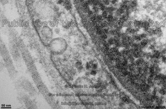
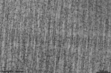
Macula adhaerens (= Desmosoma)
|
Fleckdesmosom; Verdichtungen im Interzellularraum, welche auf transmembranöse
filamentöse Proteine (Desmoglein und Desmocollin) zurückgehen.
Diese sind mit Haftplatten aus Plakoglobin verbunden, welche Ankerstellen
für verschiedene Zytoskelettkomponenten
(Keratine und andere Intermediärfilamente)
darstellen. Desmosomen halten Zellen fest mechanisch zusammen. Sie kommen
am unteren Ende von epithelialen Schlußleistenkomplexen
und besonders häufig zwischen Epithelzellen
in mehrschichtigem Epithel vor. Ferner findet man sie zwischen glatten
Muskelzellen und in Glanzstreifen
der Herzmuskulatur.
Der Durchmesser von Desmosomen beträgt 0,3 0,5 µm, der
Interzellularspalt ist in diesem Bereich 25-35 nm weit.
--> weitere Informationen und Abbildungen |
Spot-desmosome; fibrous transmembrane linker proteins (desmoglein and
desmocollin) bind to plakoglobin and other proteins in the plaques, and
extend into the intercellular space, where the fibres from the 2 cells
form an interlocking network that causes a tight connection. Bundles of
tonofilaments
including keratins are anchored to the disklike plaque, which is a mass
of electron-dense material. Desmosomes appear at the bottom of junctional
complexes of epithelial cells. Desmosomes are frequently seen between
cells of multilayered epithelium.
They are also connecting smooth muscle
cells and are included in the intercalated
disks of cardiac muscle cells.
--> further information and images |
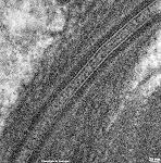
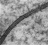
Macula communicans (= Nexus)
|
Nexus; Gap-junction, hier bilden Membranproteine durch den Interzellularraum
hindurch tunnelförmige Verbindungen, die als Konnexone bezeichnet
werden, diese bestehen aus 6 Connexin 32 Untereinheiten in hexagonaler
Anordnung. Nexus dienen der metabolischen und ionalen Koppelung benachbarter
Zellen, d.h. durch die Kanäle (Durchmesser < 1,2 nm) können
Wasser, Ionen und kleine Moleküle (< 1.500 Da), sowie cAMP von
einer Zelle zur anderen übertreten. Die rundlichen Zell-Zell Kontaktzonen
haben einen Durchmesser von 0,1 1 µm, die Zellmembranen
sind hier ca. 2 4 nm voneinander entfernt. Die Bildung erfolgt in wenigen
Sekunden. Der Öffnungszustand der Poren wird durch Ca++
und Mg++ beeinflußt.
--> weitere Informationen und Abbildungen |
Nexus, gap-junction, cell-cell contact zones 0,1 1 µm in diameter
with multiple communication channels. Cell membranes here have a distance
of only 2 4 nm. The opening of the channels is influenced by Ca++
und Mg++ concentrations and have diameters < 1.2 nm. They
allow small molecules (< 1,500 Da) water and ions to pass. Connections
are known as connexons, that consist of 6 connexin 32 molecules in hexagonal
order each. These integral membrane
proteins can form new gap junctions within seconds.
--> further information and images |
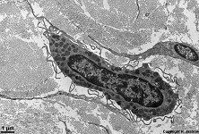
Mastocytus
|
Mastzelle; große freie
Bindegewebszelle, die sich häufig in der Nähe von Blutgefäßen
befindet. Mastzellen sollen aus aus dem Blut
ausgewanderten Stammzellen hervorgehen. Sie enthält dicht liegende
basophile Vesikel, welche Histamin und Heparin aber auch Hydrolasen Derivate
der Arachnidonsäure und Proteinasen enthalten. Die Vesikel
sind beim Menschen oft mit vielen lamellär konzentrisch geschichteten
und zylindrischen Strukturen gefüllt und von einer Membran umgeben.
Die Degranulation wird durch Immunglobulin E bei allergischen Reaktionen
vom Soforttyp hervorgerufen.
--> weitere Informationen und Abbildungen |
Mast cell; a free
cell of the connective tissue found close
to blood vessels. It shall derive from
erythropoetic germ cells and has many basophilic vesicles.
The latter contain histamin, heparin, hydrolytic enzymes and arachidon
acid derivatives. In humans the vesicles are stuffed with lamellar concentric
and cylindrical electron dense structures. Degranulation is induced by
immunglobulin E in immediate hypersensitive reactions.
--> further information and images |
| Matrix cartilaginea |
Knorpelmatrix; Knorpelgrundsubstanz, basophil und metachromatisch,
besteht aus 6070 % Wasser, Glykanen (besonders Aggrecan) 10-15 %, Hyaloronsäure,
Chondroitinsulfat, Proteoglykosaminoglykanen, Kollagen 10-15 % und Mineralien
4 %. |
cartilage matrix; basophilic and metachromatic, contains water (60-70
%), glycans (predominantly aggrecan, 10-15%), proteoglycans (10 -15 %),
collagen (10-15 %) and minerals (4 %). |
| Membrana |
Membran, mikroskopisch: dünnes elastisches Häutchen
aus Phospholipiden mit eingelagerten Proteinen, welches lichtmikroskopisch
nicht sichtbar ist und elektronenmikroskopisch als einfache oder doppelte
äußere elektronendichte Grenzschicht um Zellen bzw. deren Organellen
auftritt.
makroskopisch: dünne weiche, geschmeidige Gewebeschicht,
die Körperhöhlen auskleidet, Organe einfaßt oder voneinander
trennt. Die wichtigsten Membranen sind die Zellmembran
und die Kernmembran. |
Membrane; microscopic: thin elastic layer of phospholipids with
integrated proteins that can not be visualised in light- but only in electron
microscopy. It is a thin electron dense single or double layer limiting
cells or cell organelles.
macroscopic: a thin, soft pliable layer of tissue that lines
a tube or cavity, covers an organ or structure, or separates one part from
another. Most important membranes are the cell
membrane and the nuclear membrane |
Membrana cellularis
= Plasmalemma
|
Zellmembran = Plasmalemma = Plasmalemm.
Eine 8 nm dicke elektronenmikroskopisch aus 3 Schichten bestehende Membran:
außen 2,5 nm elektronendicht; Mitte 3 nm hell; innen 2,5 nm elektronendicht.
An Proteinen in der Innenschicht sind die Zytoskelettfilamente
verankert (Stabilität der Zelle).
Biochemisch besteht die Zellmembran aus einer Phospholipidmoleküldoppelschicht,
in die Proteine verschieblich eingebettet sind (Bioeinheitsmembran):
- als Oberflächenproteine (oft mit Zuckermolekülen gekoppelt
in der Glykokalyx, wichtig für
Blutgruppeneigenschaften,
Selbsterkennung durch das Immunsystem)
- als Tunnelproteine (durch alle Schichten reichende Ionenkanäle,
wichtig bei der Reizleitung, Koppelung von Muskelzellen).
--> weitere Informationen und Abbildungen |
Cellular membrane = plasmalemm. A 8 nm thick membrane that in the electron
microscope consists of an outer 2.5 nm electron dense-, followed by a 3
nm thick electron lucent- and an inner 2.5 nm thick electron dense layer.
Some proteins of the inner layer serve as anchors for cytosceletal
filaments (stability of the cell). Biochemically the plasmalemm is
a phospholipide molecule double layer with embedded proteins (unit membrane):
- as surface proteins (often in conjunction with sugars in glycocalyx;
important for blood group characteristics,
self-recognition of the immune system)
- as tunnel proteins (ion channels reaching through all layers, important
for electromechanicle coupling of muscle cells).
--> further information and images |
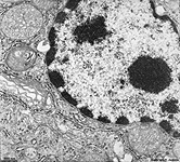
Membrana nuclearis externa
= Nucleolemma externum
|
äußere Kernmembran;
mit Ribosomen besetzte äußere
Doppelmembran des Zellkerns, die sich oft
in die Membranen von rauhem endoplasmatischen Retikulum
fortsetzt, welches sich sich in das Cytoplasma
vorschiebt. An den in die äußere Kernmembran eingelalerten Ribosomen
findet Translation von Boten Ribonucleinsäuren und damit Proteinsynthese
statt. Einige der gebildeten Proteine werden durch die äußere
Kernmembran in den von letzterer nach außen begrenzten perinucleären
Raum aufgenommen. Die Bioeinheitsmembran schließt den perinucleären
Raum und damit den Zellkernbereich nach außen ab. Sie steht an den
Kernporen
mit der inneren Kernmembran in Verbindung und ist ~8 nm dick.
--> weitere Informationen und Abbildungen
zur Kernmembran |
Outer nuclear membrane. This outer double membrane of the cell
nucleus contains ribosomes. The membrane
often continious into rough endoplasmic reticulum
streching towards the cytoplasm. Translation
of m-RNA takes place on the ribosomes of the outer nuclear membrane, resulting
proteins often diffuse through the membrane into the perinuclear space
that is bordered by the outer nuclear membrane. The outer nuclear membrane
is a unit membrane that limits the perinuclear space and has contact to
the inner nuclear membrane at the nuclear
pores. It is ~8 nm thick.
--> further information and images |
Membrana nuclearis interna
|
Innere Kernmembran; Bioeinheitsmembran,
die an die Kernlamina des
Zellkerns
direkt anschließt. Sie steht an den Kernporen
mit der äußeren Kernmembran in Verbindung und ist ~8 nm dick.
Oberhalb von ihr liegt der perinucleäre Raum.
--> weitere Informationen und Abbildungen
zur Kernmembran |
Inner nuclear membrane; unit membrane
that directly borders to the nuclear
lamina of the cell nucleus. It is connected
to the outer nuclear membrane at the nuclear pores and is ~8 nm thick.
The perinuclear space is located above it.
--> further information and images |
| Mesenchymum |
Mesenchym; embryonales Bindegewebe. Mesenchym
besteht aus noch undifferenzierten, pluripotenten mesenchymalen Stammzellen,
die sich zu Zellen des Binde- und Stützgewebes,
Muskel-,
Gefäßendothel-
und Mesothel- (also glatten Muskelzellen)
oder Zellen der Zahnanlage weiterentwickeln können. Die Zellen weisenden
viele dünne Fortsätze auf über die sie Kontakt zueinander
haben und ein Netzwerk bilden. Zwischen ihnen liegt ungeformte
Grundsubstanz. |
mesenchyme; embryonal connective tissue.
Mesenchyme consists of pluripotent still immature mesenchymal germ cells
that will become cells of connective tissue,
muscle,
endothelial
or mesothelial (i.e. smooth muscle cells)
or cells of the primordial teeth. The cells are connected to each other
via their multiple long processes forming a network. They are surrounded
by
amorphous ground substance. |
Mesophragma (Linea M)
|
M-Linie; in der Mitte der anisotropen Bande der quergestreiften Muskulatur
gelegene dunkle Linie, die beiderseits vom H-Streifen
umgeben wird. Sie entsteht durch 3 Proteinquerverbindungen zwischen den
Myosinfilamenten, dabei sind Skelemin Intermediärfilamente
beteiligt. |
M-line, dark line in the middle of the anisitropic band of striated
muscle cells surrounded on both sides by the H-zone.
It is formed by three fine filamentous structures that connect the thick
myosin filaments involving skelemin intermediate
filaments |
| Microfilamentum |
Mikrofilament = Aktinfilament
Mikrofilamente haben Durchmesser von 7-9 nm und entsprechen den Aktinfilamenten. |
Microfilament; = actin filament.
Microfilaments have diameters of 7-9 nm and are identical with actin
filaments. |
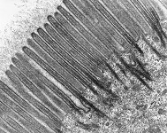
Microvillus
Abbildungen - pictures
|
Mikrovillus; feine, nur passiv wenig bewegliche, Ausstülpung der
Zellmembran mit fingerartiger Form. In der Regel treten Mikrovilli dicht
parallel zueinander gelagert in sogenannten Bürstensäumen resorbierender
Epithelzellen auf. Im Inneren der Mikrovilli befinden sich Aktinfilamentbündel,
die basal im terminalen Netzwerk verankert
sind. In der Außenmembran sind, besonders im Darm, viele Proteine
eingelagert, an denen wiederum viele Enzyme angekoppelt sind. Mikrovilli
dienen der Oberflächenvergrößerung und fördern damit
die Stoffaufnahme. Sie haben im Bürstensaum Durchmesser von ca. 100
nm und eine Länge von ca. 2 µm. Im Darm sind ihre zum Lumen
gerichteten Spitzen oft von einer hohen Glykokalix
überzogen.
--> weitere Informationen und Abbildungen |
Microvillus; a small finger-like protrusion of the cell membrane that
is not actively motile. Usually microvilli are closely stretching parallel
to each other in so called brush seams of resorbing epithelial cells. Bundles
of actin filaments are seen in the centres of microvilli. They are basal
connected to the terminal web. Lots of different proteins often connected
to enzymes are located in the outer cell membrane of microvilli. Microvilli
strongly increase the cell surface area and thus raise resorption significantly.
The diameter of a microvillus is about 100 nm and its length is ~ 2 µm.
In the gut the apex of a microvillus anchors a high glykokalix.
--> further information and images |
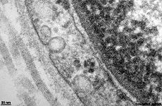
| Mitochondrion |
--> Abbildungen und Erklärungen |
--> images and explanations |
Filamentum myosinium
|
Myosinfilament, setzt sich aus Myosinmolekülen bevorzugt des Typs
I, II und V zusammen. Diese Moleküle weisen einen dickeren Kopfbereich
und einen langen Schwanz auf. Am Übergang, im Halsbereich windet sich
ein Calmodulin Molekül um das Myosin, welches im Typ 1 als Monomer,
in Typ II und V als Dimer vorliegt. Myosinfilamente bestehen aus vielen
rundlich aneinander gelagerten Myosinmolekülen, sind 325 nm lang,
wobei der mittig gelegene Myosinköfchen freie Bereich 160 nm lang
ist. Myosinköpfchen sind 16,5 x 6,5 x 4nm groß. In Gegenwart
von Aktin besitzt Myosin ATPase-Aktivität,
bei der Muskelkontraktion knicken die Myosinköpfchen um 45 Grad ab.
In mehreren Schritten ist eine Verlagerung um 11 15 nm gegenüber
den Aktinmolekülen möglich, wodurch sich die H-Linie
und die Sarkomerlänge verringert. |
myosin filament; consists of myosin molecules, mostly type I,II or
V. The latter have a thicker head (16,5 x 6,5 x 4nm ) and a long tail.
In the neck region between both a calmodulin molecule is winding around
the myosin. Myosin type 1 is a monomer, types II and V are dimers. The
myosin filaments consist of several apposed myosin molecules and have a
length of 325 nm. The middle region, which lacks myosin heads, is 160 nm
in length. When actin is present myosin
has an ATPase activity. In the muscle contraction process myosin heads
bend 45 degrees. In several steps a dislocation of 11 15 nm is possible
between actin and myosin filaments, that is the reason for shortening of
the H-line and the sarcomere. |
-->