Overview heart muscle (Textus
muscularis striatus cardiacus):
Pages with explanations are linked to the
text below the images if available! (Labelling is in German)
 |
 |
 |
 |
 |
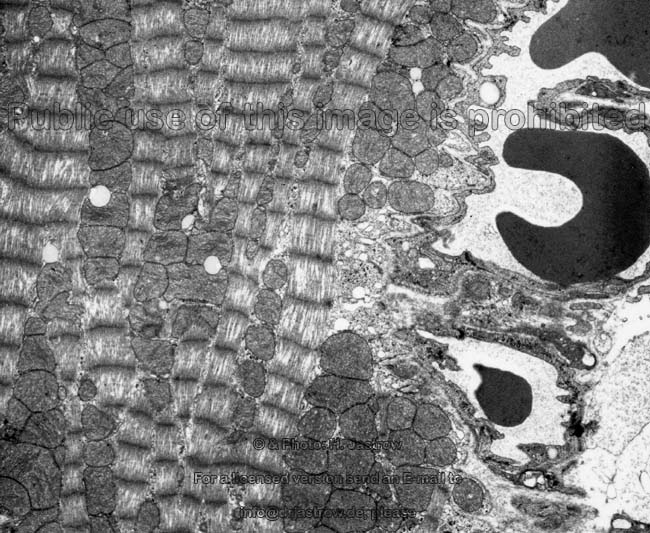
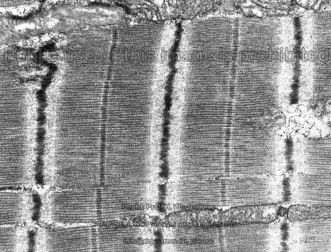
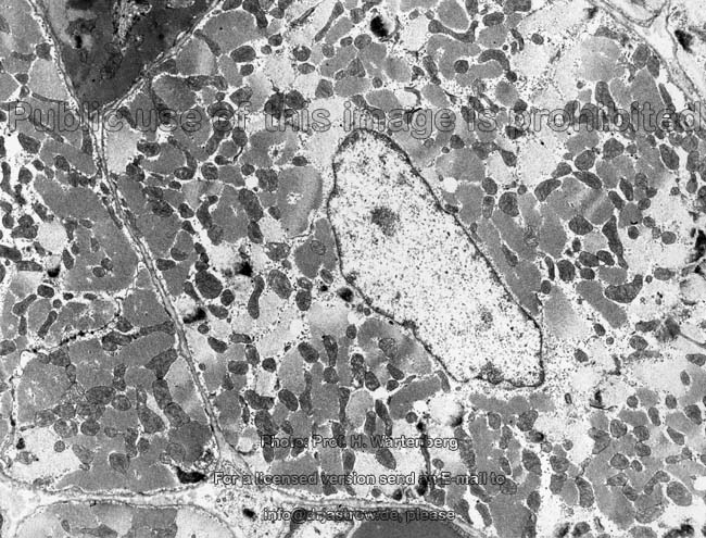
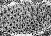
cross-section of muscle
filaments 1 (monkey) |
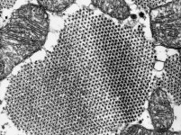
cross-section of muscle
filaments 2 (monkey) |
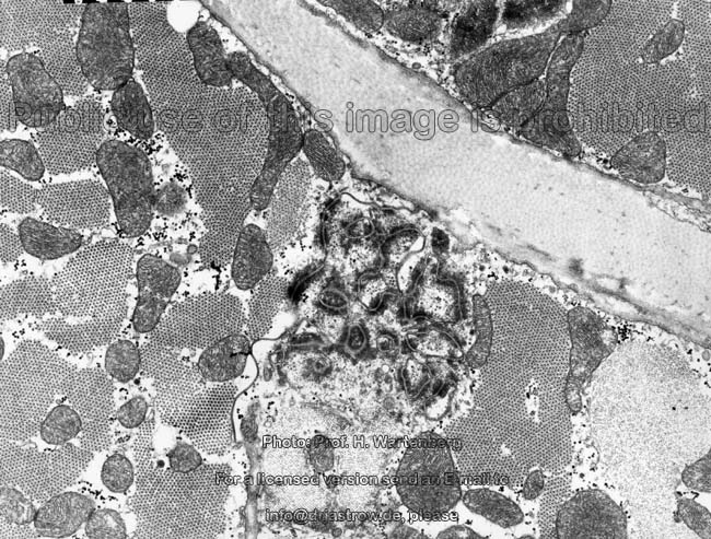
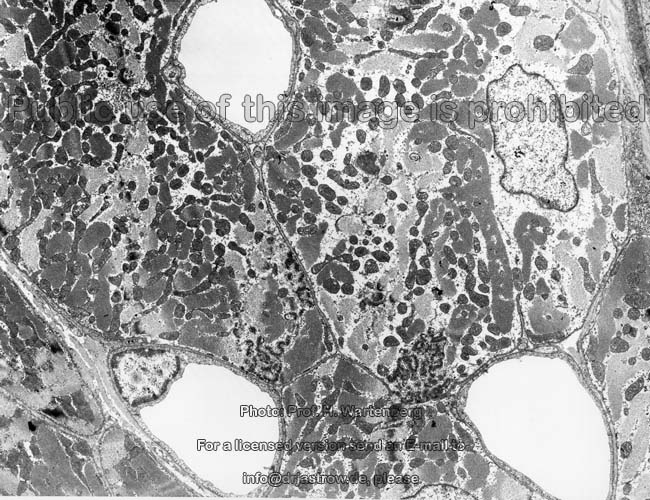
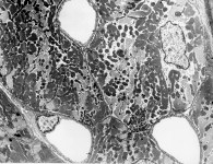
cross-sectioned overview
with capillaries (monkey) |
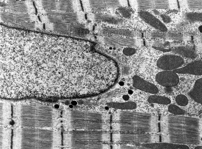
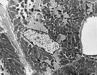
oblique section
(monkey) |
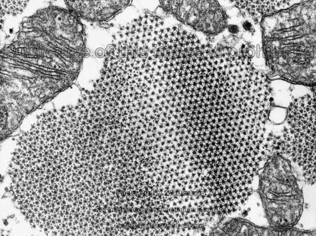
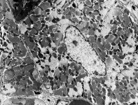
cross-section
(monkey) |
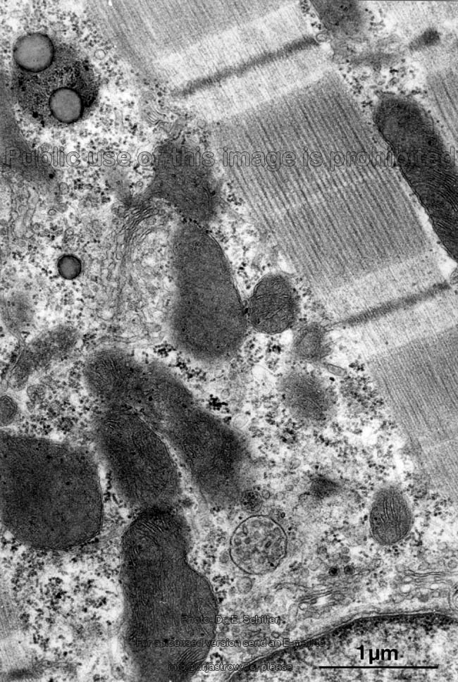
Cardiac muscle cells (Terminologia
histologica: Cardiomyocyti, Myocyti cardiaci) are about 50-150 µm
long and 10 to 20 µm thick and have a cylindrical shape. They show
typically only 1 and in some cases 2 long, central nuclei
of a cigar shape about 12 µm in length which get indented in case
of contraction. The nuclei are surrounded by less electron-dense cytoplasm
(light court; image) since myofibrils
are replaced here by the following organelles:
small
Golgi-apparatuses,
lysosomes,
lipofuscin
vesicles, and a few RER. The cells show large
amounts of mitochondria of the crista-type
with a relatively electron-dense matrix
and usually over 50 to far above 100 crests. The
arrangement of these mitochondria is characteristic,
they form large rows paralleling the myofibrils
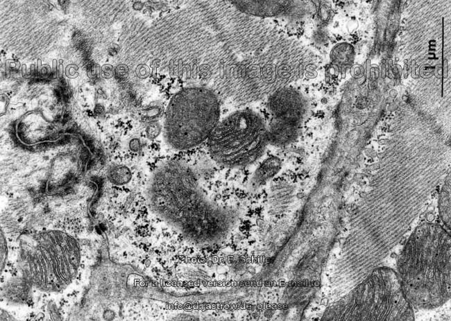
and here are called interfibrillar mitochondria. Further, only in
normal
cardiomyocytes, many
are located in groups of great number directly beneath the 9
nm thick cell membrane called sarcolemma
(Terminologia histologica: Sarcolemma). Here they are termed subsarcolemmal
mitochondria. However, the latter are in direct continuity to the interfibrillary
subpopulation on many places. The shape of mitochondria adapts to available
local space. Three-dimensionally reconstructed mitochondria
are many micrometres long, some hundred nanometres in diameter and multiple
branched tubular structures in most cases. However,
close to the nuclei they generally are more
spherical and have smaller individual volumes (perinuclear mitochondria).
One should also keep in mind that mitochondria
lack a stable inner skeleton and thus also change in shape during the muscle
contraction process. The sarcolemma shows
several micropinocytotic
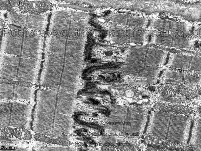
vesicles increasing in number close to capillaries. T-tubules
(Terminologia histologica: Tubuli transversi) are very deep tubular invaginations
of the cell membrane in regions of Z-discs
as can be seen here. They always run along
the outer edge of each Z-disc
in a distance of only a few nanometres surround the discs and then continue
to Z-disc of the neighbouring
parallel running myofibril. Thus the T-tubules
are in direct continuity with the extracellular space and every sarcomere
is supplied by only one such tubule whereby neighbouring tubules are interconnected
by longitudinal tubules. In comparison to the skeletal
muscle cardiac T-tubules are a little wider (diameter 100 - 300 nm
in man; considerably thinner in mouse and rat). Thus the basal
lamina also enters the T-tubules as intratubular basal lamina
(Terminologia histologica: Lamina basalis intratubularis). The latter has
a little thinner lamina densa
which in parts seems to be interrupted. It
is anchored to the cell membrane by alpha5-beta1-integrins.
The latter is stabilised by cytoskeletal
elements like actin, vinculin and spectrin
and has many integrated voltage-dependent channels for calcium ions (Ca++,
DHP-receptors). This is of functional importance since thus extracellular
Ca++ may easily and quickly penetrate deeply into the cell via
the T-tubules.
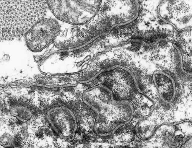
The smooth endoplasmic reticulum
of cardiac muscle cells is termed sarcoplasmic reticulum (SER;
Terminologia histologica: Reticulum sarcoplasmicum). In the heart it is
not as prominent as in skeletal muscle and
forms a network of multiply interconnected, longitudinally oriented to
the orientation of the myofibrils, therefore
the term L-tubules. The tubules partly have tubular elements
(Terminologia histologica: Elementa tubularia) which are connected to reticular
elements (Terminologia histologica: Elementa reticularia), i.e., net-like
interconnected tubules. At the level of the M-line
the network of L-tubules is denser and it stretches towards the T-tubules
up to a distance of only few nanometres showing terminal
cisterns (junctional SER; Terminologia histologica: Cisternae terminales)
which are local dilatations about 70 nm in diameter. In
the sections in which one such cistern is facing a T-tubule
for several hundred nanometres the resulting complex is termed a diad
(Terminologia histologica: Dias) since when this is cross-sectioned one
will see two parallel lying tubes. These diads are only seen in
heart- but NOT in skeletal muscle. However,
often two terminal cisterns of the SER also accompany
one T-tubule, which then is in the centre between
the two cisterns which results in formation of a triad (Terminologia
histologica: Trias) with 3 parallel tubes. On
some places the SER nearly attaches to the sarcolemma
while corbules, i.e. vesicular protrusions (corbular SER; missing
in Terminologia histologica; proposal: Corbulae). The SER is the most important
short term storage for calcium ions (Ca++), whereby
especially the terminal cisterns and the corbular SER with the Ca++-binding
protein calsequestrin and the
receptors present in the SER membranes (ryanodin receptor in the terminal
cisterns or a IP3-regulated receptor in the corbular SER, respectively)
are of importance. In order to realise a quick relaxation of the heart
muscle after contraction which is important for refill of the ventricles
during the diastole ATP-consuming
calcium
ion pumps are located in the membranes of the SER. These
pumps are regulated by the cardiac protein phospholamban and rapidly
pump the Ca++ back into the lumen
of the SER. Further calcium ion pumps regulated
by Calmodulin are present in the cell membrane
and in the membrane of the T-tubules in order to
quickly pump back Ca++.
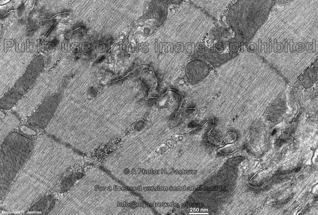
The complex intercellular junctions
(Terminologia histologica: Junctiones intercellulares
speciales) connecting heart muscle cells consist
of interdigitations plus adhesive strips
(Fasciae adhaerentes),
spot desmosomes (Terminologia
histologica: Maculae adhaerentes) and gap-junctions
(Terminologia histologica: Maculae communicantes,
Nexus).
--> Differential diagnosis of muscle tissues,
skeletal
muscle, cell-to-cell junctions, caveolae,
L-tubules,
actin
filaments
--> Electron microscopic atlas Overview
--> Homepage of the workshop
Some images were kindly provided by Prof. H. Wartenberg;
other images, page & copyright H. Jastrow.