Overview blood cells:
Pages with explanations are linked to the
text below the images if available! (Labelling is in German)
| Names |
red blood cells
(erythrocytes)
|
w h i t e
b l o o d c e l l s ( l e u k o c y t e s )
|
platelets
(thrombocytes)
|
|
granulocytes
|
lymphocytes |
monocytes |
|
neutrophil
|
eosinophil
|
basophil
|
|
with rod-like nuclei
|
with segmented nuclei
|
example
images |
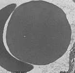
|
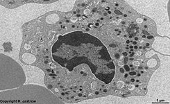
|
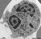
|
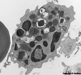
|
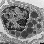
|
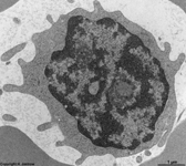
|
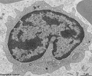
|
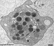
|
number per
1 litre of blood |
m: 4.4 - 6.0 x 10¹²
w: 3.8 - 5.2 x 10¹² |
0,52 x 10⁹ (mean) |
3,0 x 10⁹ (mean) |
0,15 x 10⁹ (mean) |
0,03 x 10⁹ (mean) |
2,5 x 10⁹ (mean) |
0,43 x 10⁹ (mean) |
250 x 109 (mean)
150 - 400 x 109 |
frequency in differential
white blood count |
- |
9,5 % (mean) |
40,5 % (mean) |
3,2 % (mean)
0 - 7 % |
0,6 % (mean)
0 - 2 % |
36,0 % (mean)
20 - 50 % |
7,1 % (mean)
3 - 9 % |
- |
| diameter |
7,5 µm (mean) |
9-12 µm |
9-12 µm (mean) |
11 - 14 µm |
8 - 11 µm |
7 - 10 µm |
12 - 20 µm |
1 - 4 µm |
average single
cell volume |
82 - 96 µm³ (femtolitres) |
ca. 320 µm³ (femtolitres) |
ca. 320 µm³ (femtolitres) |
ca. 400 µm³ (femtolitres) |
ca. 270 µm³ (femtolitres) |
230 µm³ (femtolitres) |
470 µm³ (femtolitres) |
6 µm³ (femtolitres) |
The table shows all major blood cells (Terminologia histologica:
Haemocyti) which make up about 45% of the blood volume. This value
(percentage of blood cells from blood volume) is called hematocrit
and is 50-60 in new-borns, 30-40 in small children and 40-50 in human adults.
Red
blood cells (erythrocytes; Terminologia
histologica: Erythrocyti, Haematiae) are distinguished from
white blood
cells (Leucocytes; Terminologia histologica: Leucocyti) and
platelets
(thrombocytes; Terminologia histologica:
Thrombocyti) which are no complete cells but fragments segregated from
megacaryocytes.
Leucocytes
comprise the following cells: lymphocytes
(Terminologia histologica: Lymphocyti), monocytes
(Terminologia histologica: Monocyti), neutrophilic
granulocytes (neutrophils, segmented neutrophilic granulocytes;
Terminologia histologica: Granulocyti neutrophili, Neutrophili, Granulocyti
neutrophili segmentonucleares), eosinophilic granulocytes
(eosinophils; Terminologia histologica: Granulocyti acidophili, Eosinophili)
and basophilic granulocytes (basophils; Terminologia histologica: Granulocyti
basophili, Basophili). Besides the cells mentioned before about 22 - 139
reticulocytes
(Terminologia histologica: Reticulocyti) precursors of erythrocytes
still containing small numbers of cell organells like mitochondria,
Golgi-vesicles
or lysosomes) are present per nl (0,9 - 2,3
% in differential white blood count). Further, considerably less plasma
cells and hardly any precursors of leucocytes or hematopoetic
stem cells are seen in normal blood.
Blood cell formation takes place in the red
bone marrow under normal conditions. However, during ontogenesis, i.e.
in embryos it primarily occurs in the yolk sac then it migrates into liver
and spleen. As soon as the bones
begin with chondral ossification in fetuses blood cell formation begins
to completely move into the bone marrow. Finally,
under normal conditions, the red bone marrow
takes over the entire blood cell
formation.
Blood cells are transported and circulated through the body in blood
vessels. However, all leukocytes are capable to leave the vessels and
to migrate into differnt kinds of connective tissue
as well as into several epithelia.
The blood plasma (Terminologia histologica:
Plasma sanguinis) is the fluid, cell-free portion of the blood which makes
up about 54 - 56% of the blood volume. It contains 6.5 to 8% of
proteins and 1 % of other components like hormones, lipoids, sugars, electrolytes
and vitamins. Fibrinogen (haemostasis
factor I), a 340 KD elongate glycoprotein of 6 polypeptid chains comprises
about 2.5% of the entire plasma proteins and has a value of 3 g/L. Prothrombin
a vitamin K-dependent protease deriving from hepatocytes (72 KDa) has a
concentration of 0,1-0,15 g/L in plasma. The
colloidosmotic
pressure of blood plasma usually is 25 mm Hg.
If the blood plasma is deprived from the coagulation
factors and substances involved in coagulation it is called blood serum
(missing in Terminologia histologica; proposal: Serum sanguinis). In other
words this is the remaining fluid after clotting.
--> Blood vessels, Blood
barriers
--> Electron microscopic atlas Overview
--> Homepage of the workshop
Images, page & copyright H. Jastrow.


