Overview Microvilli (Microvilli):
Pages with explanations are linked to the
text below the images when available
 |
 |
 |
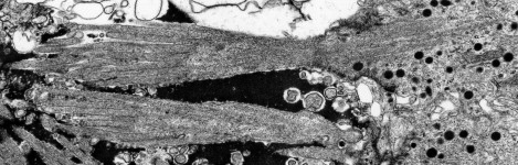 |
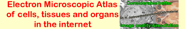
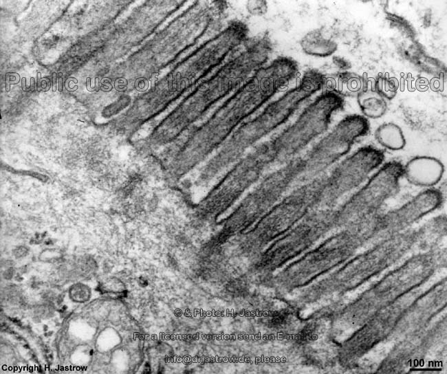
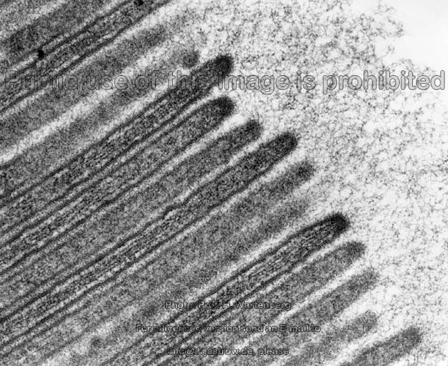
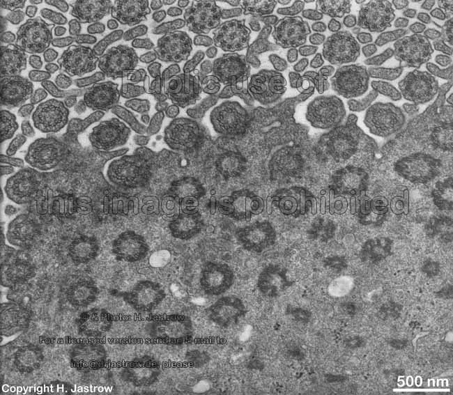
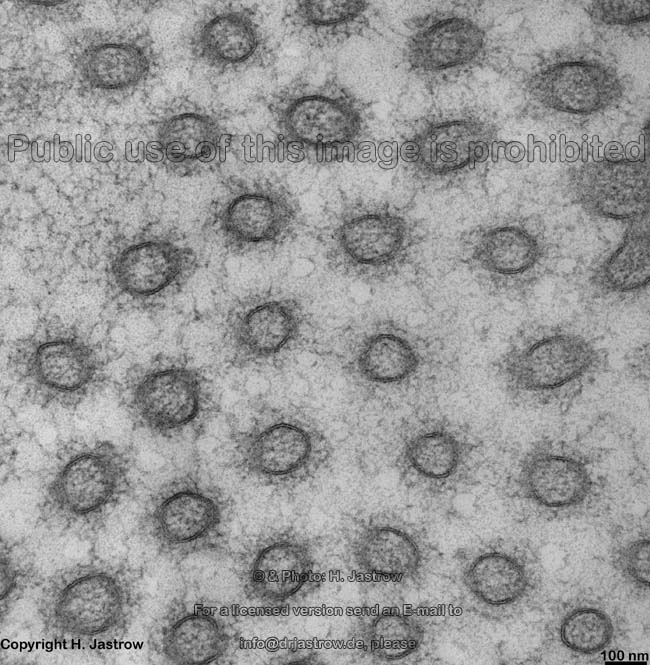
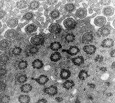
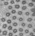
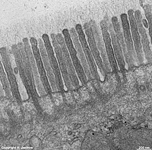
microvilli kidney
proximal tubule (monkey) |
microvilli kidney proxi-
mal tubule (monkey) 2 |
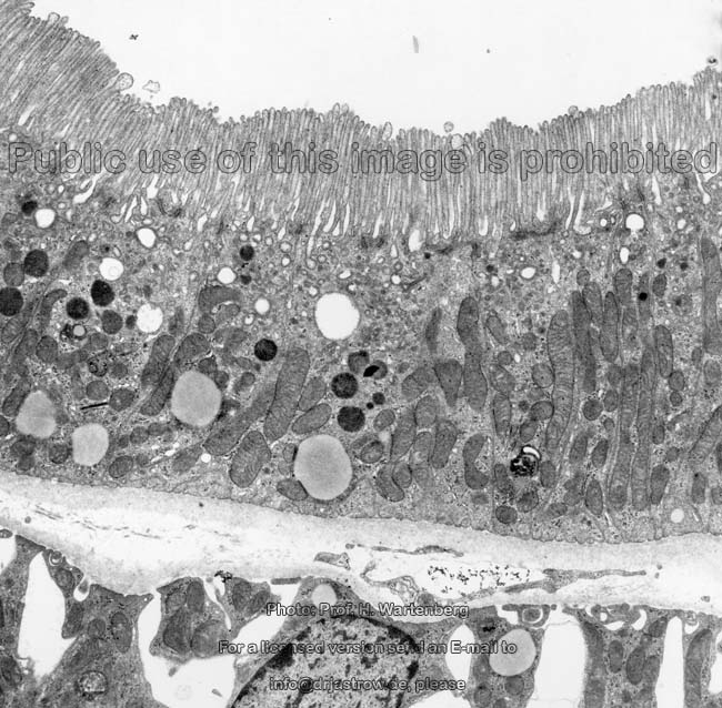
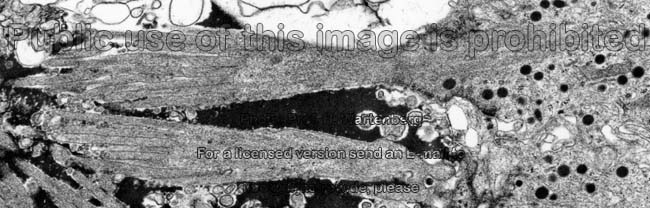
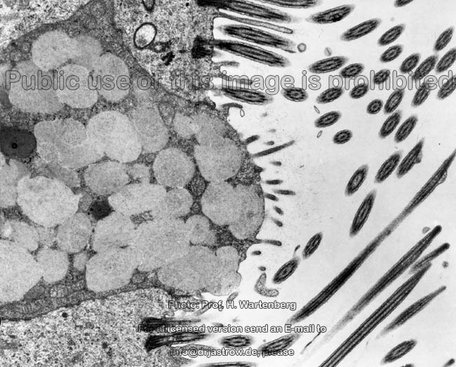
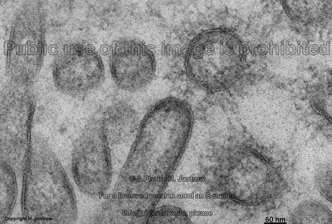
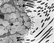
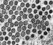
microvilli + Acinus
(exokrine pancreas, monkey) |
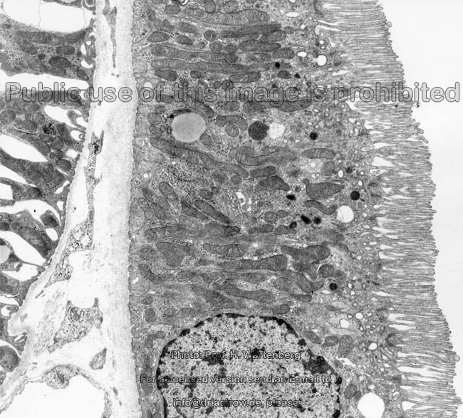
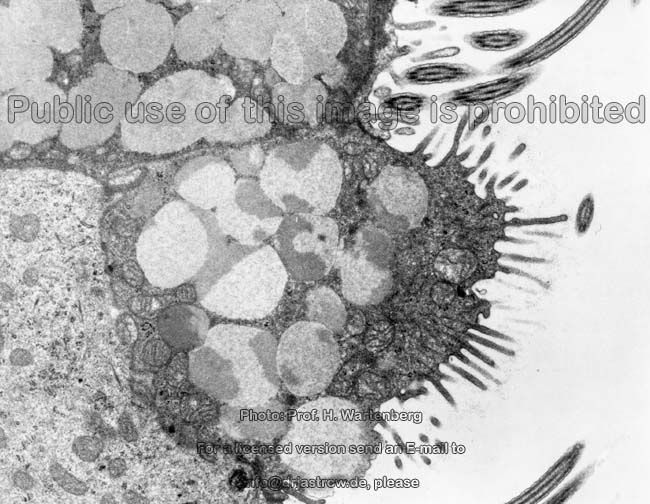
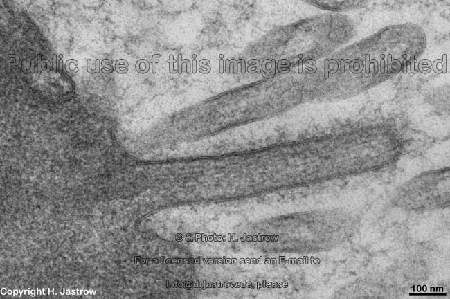
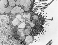
sensory microvilli of
a taste pore
(monkey) |
 |
 |
 |
 |
 |
 |
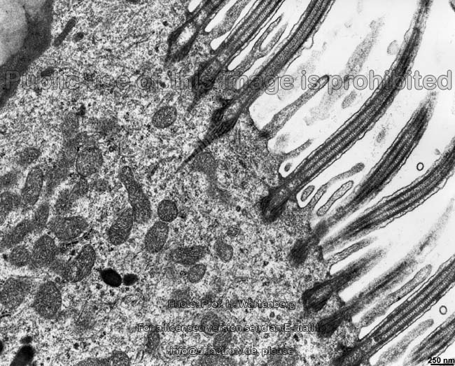
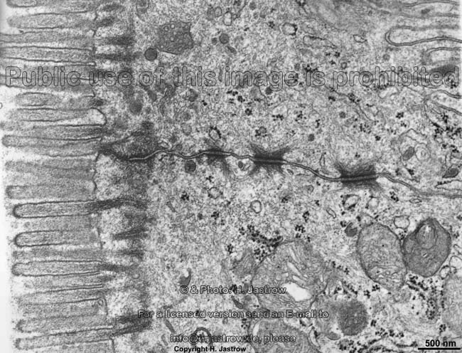
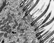
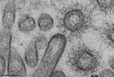
microvilli goblet cell + Ki-
nocilia, trachea (monkey) |
microvilli + kinocilia
(trachea, monkey) 2 |
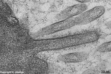
kinocilia and
microvilli 3 (monkey) |
Kinocilia + Micro-
villi cross section (monkey) |
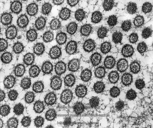
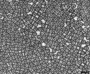
Microvilli cross section
with glycocalyx (monkey) |
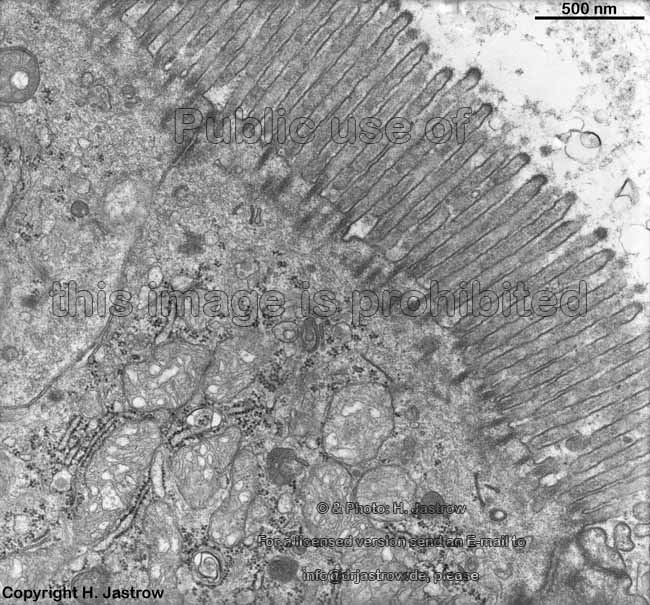
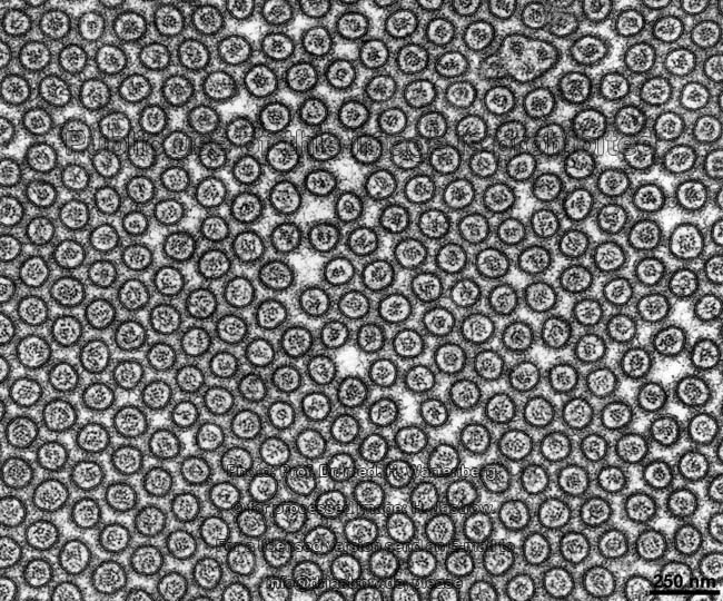
Duodenum: Microvilli
cross section (monkey) |
 |
 |
 |
 |
 |
 |
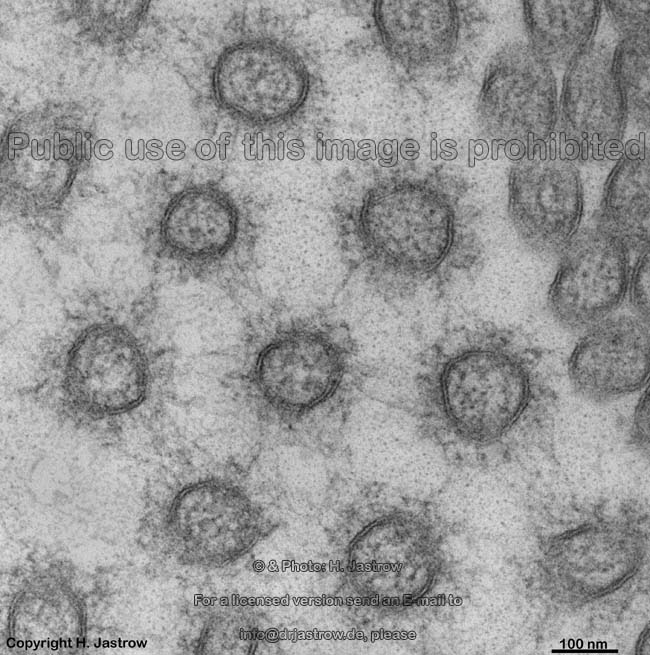
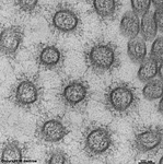
microvilli + glycocalix 1
(human pharyngeal tonsil) |
idem overview |
microvilli + glycocalix 2
(human pharyngeal tonsil) |
microvilli + glycocalix 3
(human pharyngeal tonsil) |
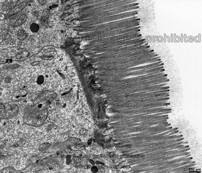
microvilli of the gut
(rat) |
microvilli of the colon +
large glycocalix (rat) |
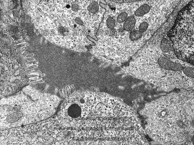
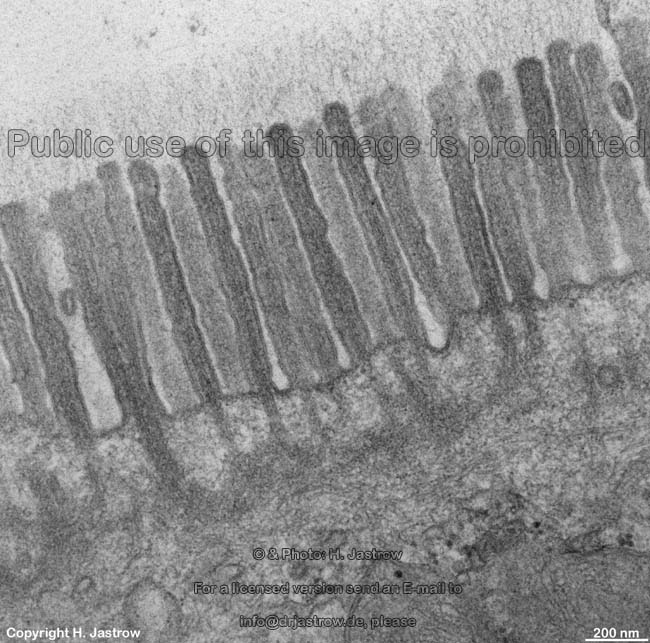
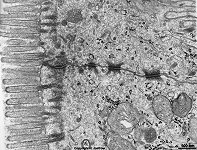
Microvilli (Terminologia anatomica: Mikrovilli) are small
finger-like protrusions of the cell
membrane towards a lumen that
are not actively motile. In many
cases microvilli form brush borders consisting of regularly ordered
microvilli closely stretching parallel to each other on the surfaces of
resorbing
epithelial cells. The diameter
of a microvillus in a brush border of intestinal
cells is about 50 - 100 nm and its length is 1 - 2
µm, however, in other epithelial cells
length is about 100 - 200 nm and the diameter is about 50 nm. Bundles of
20 to 40 actin filaments embedded in
cytoplasm are present in inside of the
microvilli. They are basally connected to the terminal
web. Apically the filaments are attached to the thickened cell
membrane on top of the microvilli where they are anchored
by myosin 1. The
actin
filaments are bundled with help of fimbrin
and
villin that also keep them
in distance to each other. In the terminal
web the actin filaments interact with spectrin
and myosin-2 filaments which is supposed to
cause smallest movements in order to support resorption of substances from
the lumen of e.g., gut. Microvilli
strongly increase the cell surface area - about 600 -
times and thus raise resorption significantly. Thus the active surface
for resorption in an adult gut altogether
raises to about 200 m². Lots of different proteins often connected
to enzymes are present in the outer membrane of the microvilli which
comprises the cell membrane, especially
in the gastrointestinal tract. For example
lots of different ion channels, amino acid transporters, transporters for
glucose and many other substrates. Thus the vast majority of substances
which are resorbed in the gastrointestinal
tract is taken up into the cytoplasm
the area of the microvilli though this cannot be visualised by conventional
electron microscopy.
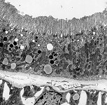
Pinocytosis,
an uptake (endocytosis) with formation
of vesicles, i.e. macropinosomes
is only possible in the small areas of the cell
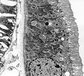
membrane located in between the bases of adjacent microvilli. The calcium
ion-binding proteins villin and gelsolin
are present in the tip regions of microvilli. They may induce a local destruction
of the cytoskeleton caused
by calcium ion influx e.g., in renal tubules
which is followed by an inspecific
apocytosis with release of small vesicles into the tubular lumen. In
the gut the apex of a microvillus anchors
plenty of small interconnected filaments, the antennulae microvillares.
The latter form the glycocalyx.
--> further cell surface differentiations:
kinocilia,
stereocilia
--> Electron microscopic atlas Overview
--> Homepage of the workshop
Some images were kindly provided by Prof. H. Wartenberg;
other images, page & copyright H. Jastrow.