List of abbreviations
Vocabulary
of micros-
copic
anatomy
specialist terms
explained in
English +
German

Every attempt was made to provide correct information and labelling, however any liability for eventual errors or incompleteness is rejected!

dieser Seite

Editor:
Dr. med.
H. Jastrow


Conditions
of use
Overview arteries (Arteriae):
Pages with explanations are linked to the text below the images if available! (Labelling is in German)
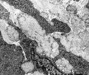
Aorta Tunica media
(monkey)
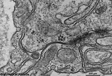
umbilical cord artery
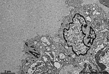
endothelium (human)
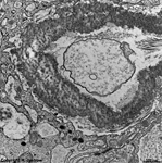
umbilical cord artery
endothelial cell 1 (human)
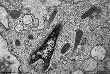
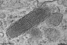
Detail: Waibel-Pallade
body (human)
umbilical cord artery en-
dothelial cell 2 (human)
Detail: junctional
complex (human)
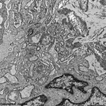
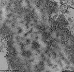
elastic membrane um-
bilical cord artery (human)
Detail: elastic
membrane (human)
arteries (pulse vessels; Terminologia histologica: Arteriae) are large vessels that carry blood away from the heart. Ther are two kind of arteries: those of the elastic type (large arteries close to the heart: aorta, pulmonary trunc, brachiocephalic trunc, common- internal- and external carotid artery, vertebral artery, common- external and internal iliac artery, femoral artery and subclavian artery) and those of the muscular type (all other arteries).
 The wall of an artery has the following layers when looking form internal to external:
1. Tunica intima consists of endothelium, the Stratum subendotheliale and the internal elastic membrane (Membrana elastica interna).
The endothelium is a monolayer of squamous endothelial cells (The squamous character of the endothelial cells can only be seen under the high pressure physiologically present in living individuals. Since preparations are usually NOT fixed and embedded under pressure endothelial cells artificially appear cuboid or even columnar in most preparations).
The stratum subendotheliale (Lamina propria intimae) is a small loose connective tissue layer with fine elastic webs and intermingeled smooth muscle cells. This layer is well developed in arteries of the elastic type but lacks in smaller arteries of the muscular type.
The internal elastic membrane (Membrana elastica interna), is a rather thick membrane of elastic fibres. It is the border to the media and easily detectable only in arteries of the muscular type.
2. Tunica media The media is comprised of larger lamells of elastic and less collagenous fibres in web- like interconnection with scattered smooth muscle cells. While in arteries of the elastic type elastic fibres clearly dominate, arteries of the muscular type show considerably larger amounts of smooth muscle cells.
3. Tunica externa (adventitia) Its external elastic membrane (Membrana elastica externa) is the border to the meda and is obvious only in arteries of the muscular type. The outer adventitia consists of mainly loose connective tissue with many fibrocytes and free cells. The adventitia serves for anchoring the artery to surrounding tissues and contains the blood vessels responsible for nutrition of the artery (Vasa vasorum) as well as non-myelinated nerve fibres.

--> arteriols, blood vessels, endothelial cells
--> Electron microscopic atlas Overview
--> Homepage of the workshop


One images was kindly provided by Prof. H. Wartenberg; other images, page & copyright H. Jastrow