Fig.1
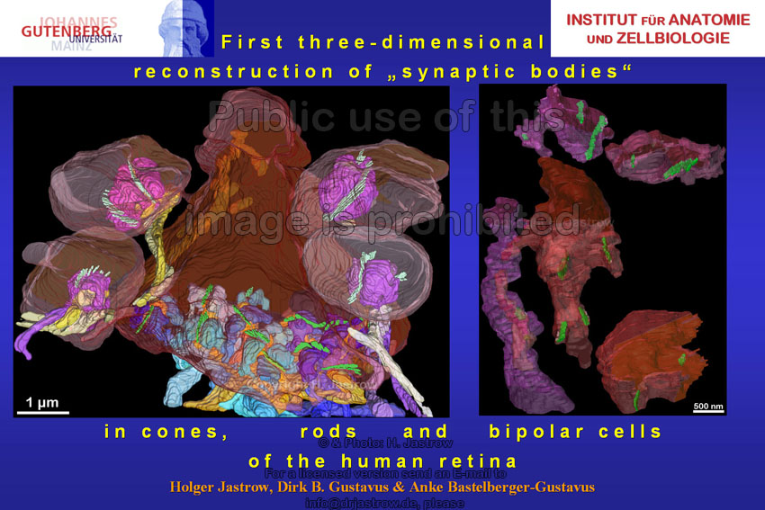 |
The retina is the most important sensory area in mankind. Its general structure and organization is well known today. However, still few attempts have been made to elucidate the ultrastructural morphology of the first and second synapses of the visual pathway which are located in the plexiform layers (Fig.2). | Fig.2
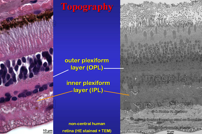 |
Fig.3
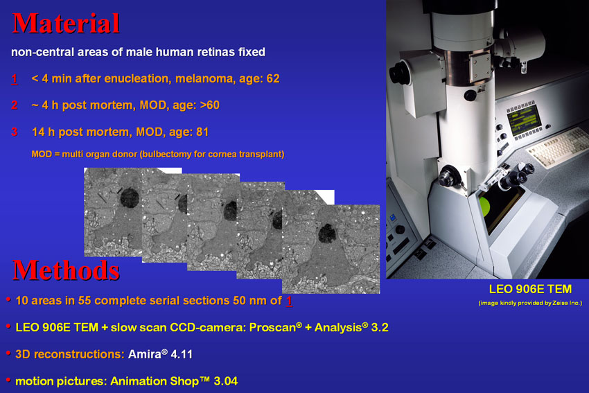 |
The tumour-free half of an enucleated eye was kindly provided by a 62-year-old malignant melanoma patient.
Only this immediately fixed material was of suitable quality for reconstruction and visualisation using the mentioned hard- and software
(Fig.3).
The high activity synapses of rods and cones are characterized by pre-synaptic bodies anchored to the cell membrane by arciform densities and invaginations of their postsynaptic elements (Fig.4). These are rod or cone bipolar cell dendrites (shown in yellow to orange) and horizontal cell processes (in blue to purple). As we will see soon most of the vesicle-binding electron-dense organelles appear somewhat ribbon-like in 3D. For this reason the term synaptic ribbons has been established. |
Fig.4
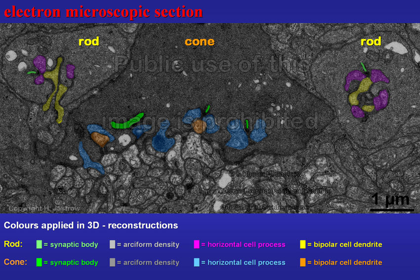 |
Fig.5
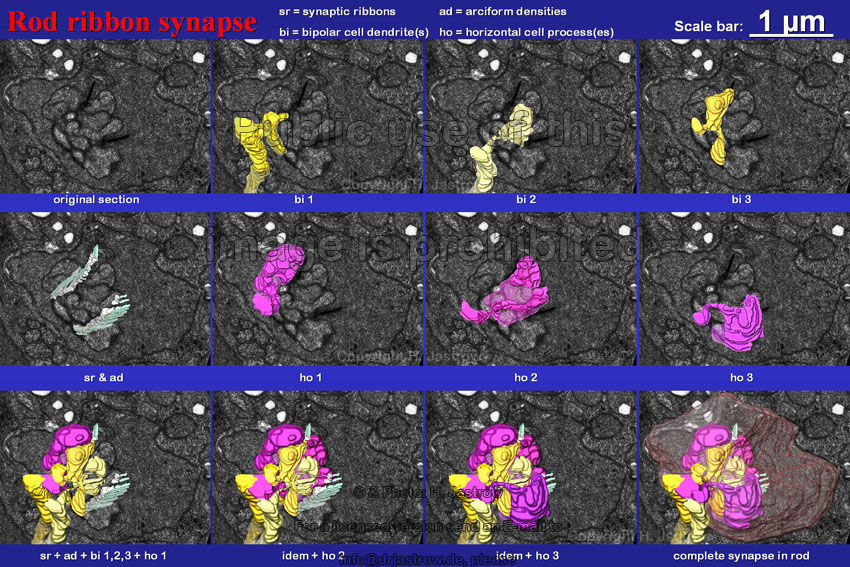 |
Fig.5 shows an example of a rod ribbon synapse. It is comprised of two synaptic ribbons with arciform densities,
three centrally located bipolar cell dendrites, and three horizontal cell processes. The bend and branched postsynaptic processes irregularly wind around
each other and interfere with small finger-like protrusions of the rod. This results in a rather complicated arrangement of the complete synapse.
The stereo animation (Fig.6) shows another rod terminal with three ribbons, three horizontal and four central bipolar cell processes. Please note that all these processes enter the terminal in a very tiny pore. Perhaps you can imagine the finger-like protrusions of the rod cytoplasm which is not visualized here. |
Fig.6 -
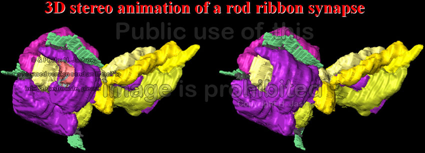 |
Fig.7
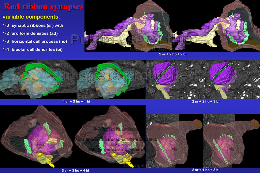 |
Reconstructions of several different rod ribbon synapses (Fig.7) demonstrate that synaptic ribbons typically lie on top of the central bipolar cell dendrites and are laterally flanked by horizontal cell processes, but that there is considerable variation regarding morphology and quantity of these components. These stereo images of five different rod synapses show one up to four bipolar cell dendrites and one to three horizontal cell processes. There may be one, two or three synaptic ribbons. Which may be connected to the cell membrane via one or in a few cases two different arciform densities. Most commonly two synaptic ribbons lie opposite to each other. These cell organelles may have variable shapes as we see in Fig.8 on the example of 11 different ribbons of rods shown in sagittal, transverse and frontal orientation. Whereas the thickness of the organelles is constant at 35 nm, their parallel major flat surfaces show considerable variations. Most structures are somewhat ribbon-like but a few are strongly bend in one plane resulting in C-, U- or V-like forms. | Fig.8
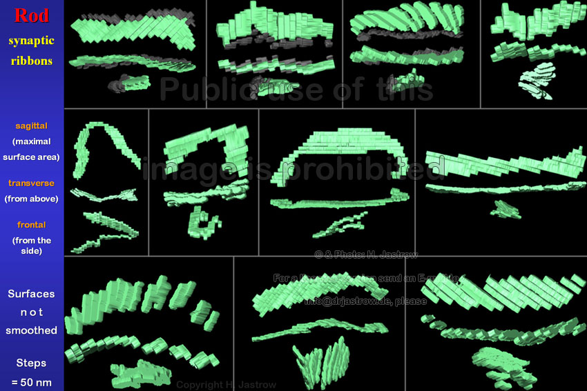 |
Fig.9
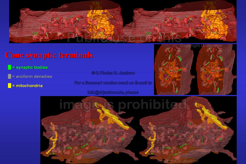 |
Even with series of 55 sections only parts of cone synaptic terminals could be reconstructed. Please note that ribbon synapses are only present in the dip of the centre of the three different cone terminals shown in Fig.9. The reconstructed tissue bloc of Fig.10 represents less than half of a total cone terminal. Such terminals, which have diameters of 5 to over 10 micrometers, are considerably larger than rod terminals. Interestingly, mitochondria are grouped in lateral areas whereas the centre of this terminal virtually only shows vast amounts of synaptic vesicles. Some of which are bound to the 12 synaptic ribbons. Be aware that only the cell processes involved in the ribbon synapses are shown here and please note the processes to nearby rods in the upper area. | Fig.10
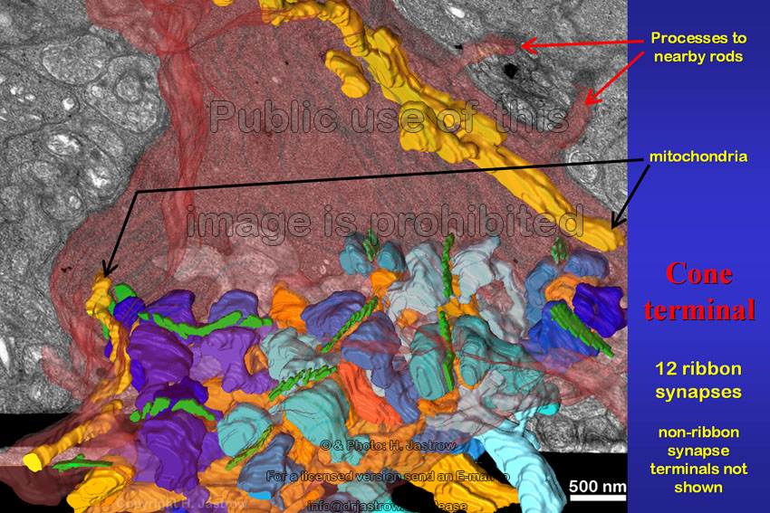 |
Fig.11
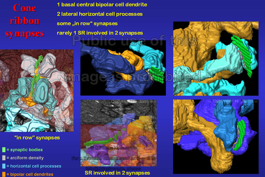 |
A closer look at some examples of cone ribbon synapses (Fig.11) demonstrates that they are simpler than rod synapses
with relatively straight, much shorter invaginations of a thick bipolar cell dendrite inferior to the ribbon and two flanking horizontal cell processes.
In some cases two rarely three ribbons are in row to each other on one and the same bipolar cell dendrite. Eventually, one single synaptic ribbon may be involved
in two different synapses. Fig.12 prooves that there may be even more than six different terminals of branching bipolar cell dendrites each of which may be associated to one or two synaptic ribbons. Further two parallel ribbons may be related to one identical horizontal cell processes as you see on the right or they may lie quite close to such of neighbouring synapses (lower left). |
Fig.12
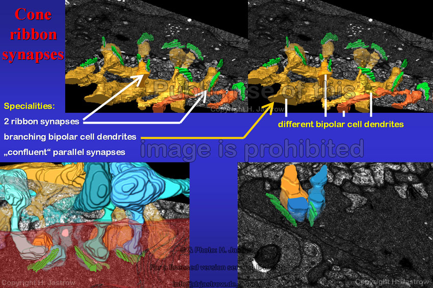 |
Fig.13 -
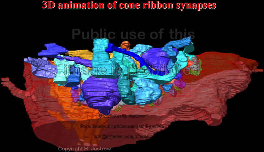 |
The animation (Fig.13) demonstrates 12 ribbon synapses with their horizontal and bipolar cell processes which intermingle and branch in the outer plexiform layer. The small lateral processes of the cone itself contact similar processes of neighbouring cones. Application of 3D reconstruction reveals that synaptic bodies of cones (Fig.14) are predominantly ribbons. However a considerable number of the organelles is slightly curved and appears like boomerangs. On an average these organelles are a little smaller in cones than in rods and have even fewer torsions. | Fig.14
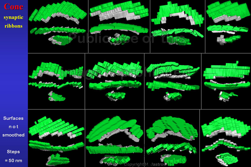 |
Fig.15
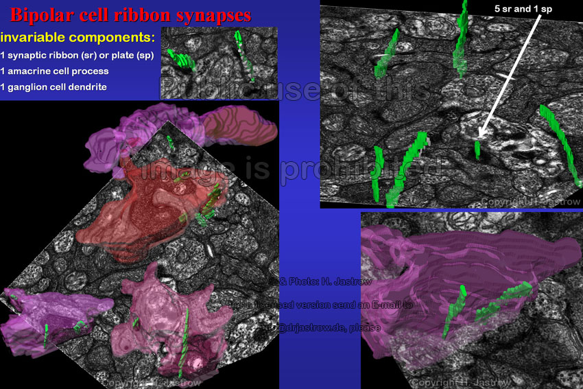 |
The synapses of the bipolar cell axons in the inner plexiforme layer are nearly flat and show only one single synaptic ribbon or small plate opposite to the border of the postsynaptic ganglion cell dendrite and amacrine cell process (Fig.15). In most, but not all cases a small electron-dense anchoring material attaches the ribbon to the cell membrane. The animation (Fig.16) demonstrates 14 synaptic bodies of five different bipolar cell axons in the inner plexiform layer. Note that some of them are small plates and that attachment plaques are shorter and in some cases lacking. | Fig.16 -
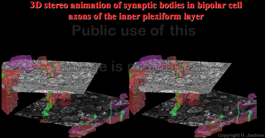 |
Fig.17
 |
Only here in bipolar cells small plate-like synaptic bodies with width to length ratios less than 1 : 3 are present whereas
most of them are quite long ribbons with little bends or torsions (Fig.17). Fig.18 summarises most important findings on retinal ribbon synapses and synaptic bodies in humans. The complicated arrangement especially of rod ribbon synapses and the vast number of postsynaptic processes in cone terminals explain why it is so difficult to understand what really happens during signal transduction in the visual system. |
Fig.18
 |
Fig.19
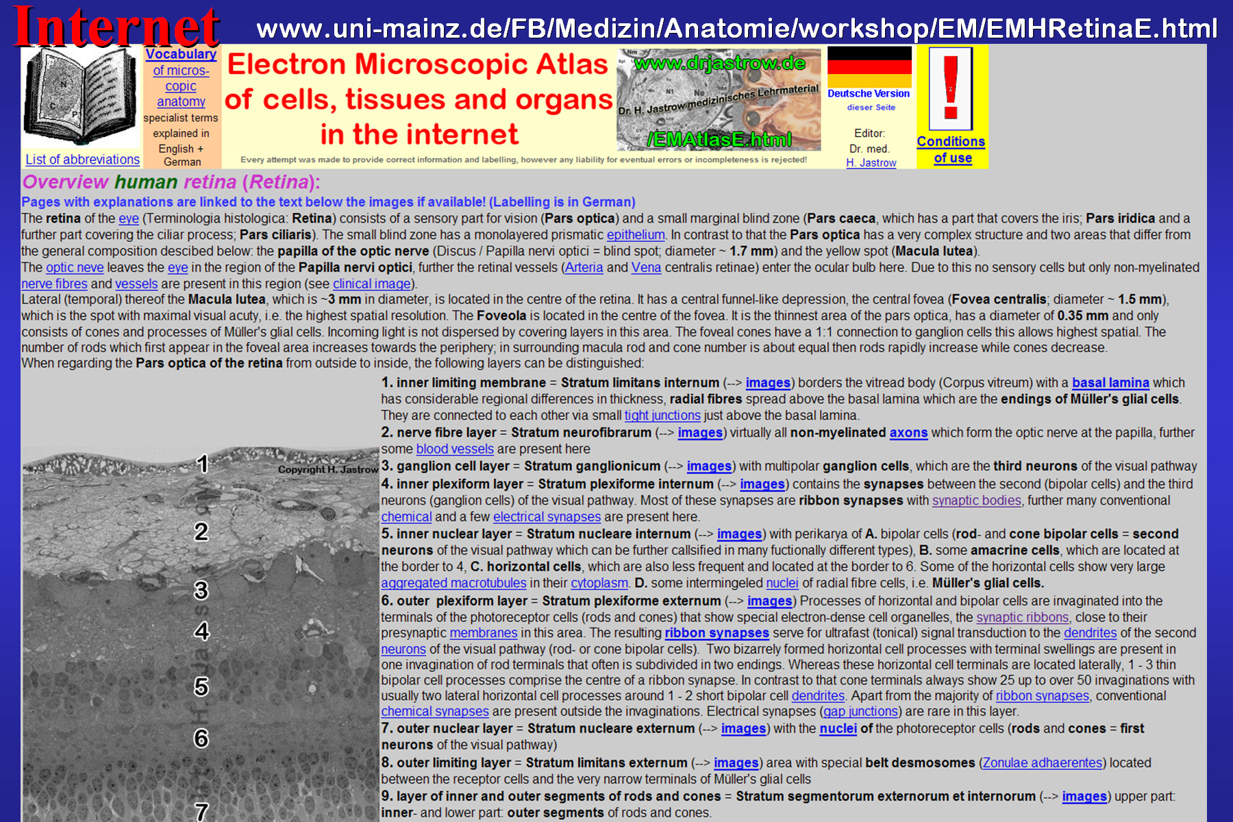 |
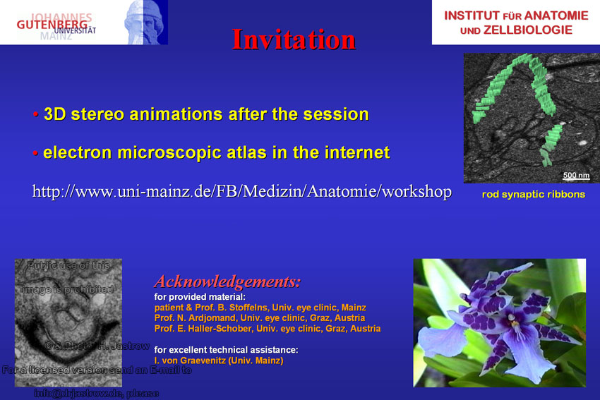
More information about the human retina may be retrieved in the electron microscopic atlas in the internet on
http://www.uni-mainz.de/FB/Medizin/Anatomie/workshop/EM/EMHRetinaE.html (Fig.19). We are grateful to the persons mentioned on Fig.20 for their support and, finally, with the smiling ribbon thank you for your kind attention. |
Fig.20
 |