Overview pigment cells (Cellulae
pigmenti):
Pages with explanations are linked to the
text below the images if available! (Labelling is in German)
|
|
 |
 |
 |
 |
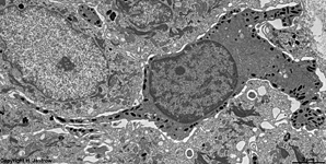
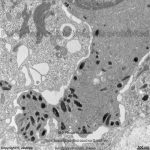
pigment epithelium of the iris
(monkey) |
melanocyte with a long process
human skin |
detail therof
basal process |
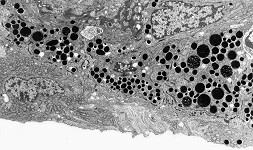
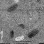
melanosom 1
human skin |
melanosom 2
human skin |
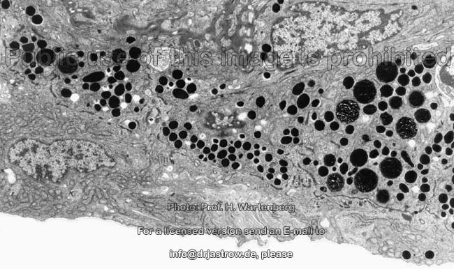
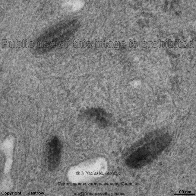
phagocyted melanosomes in a
keratinocyte from human skin |

|
|

|
|
|
|
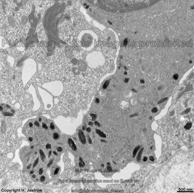
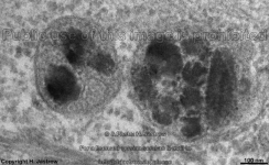
melanosomes 1
human melanocyte |
melanosomes 2
human melanocyte |
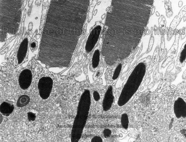
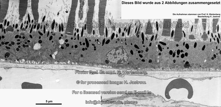
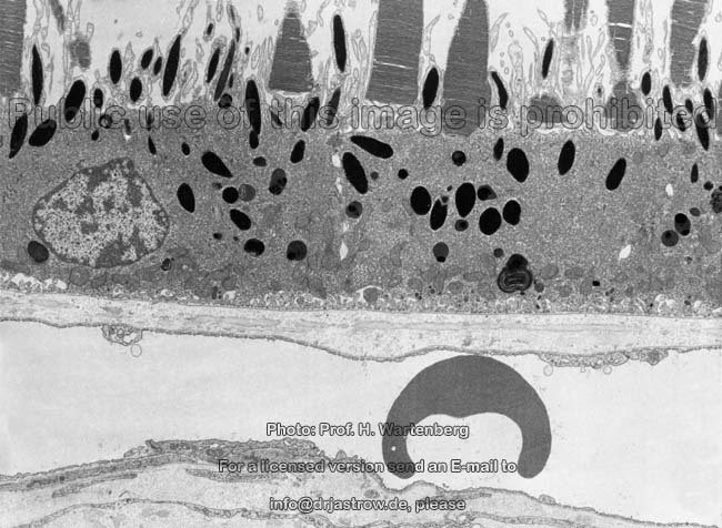
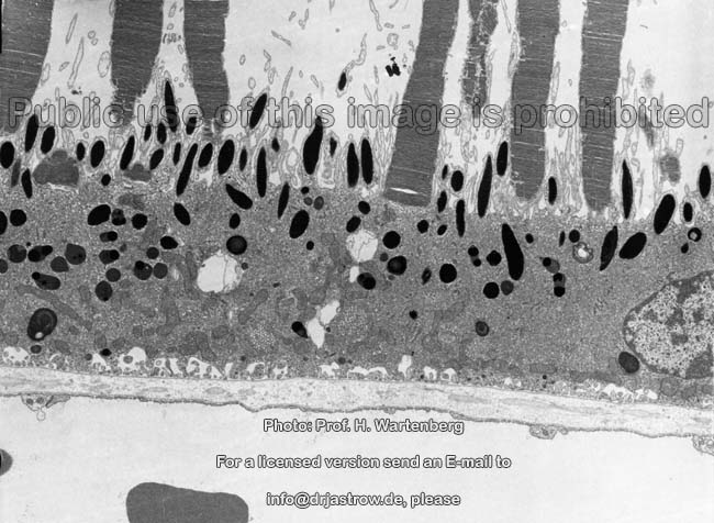
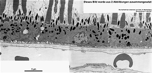
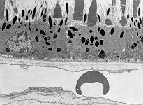
pigment epithelium + rod
outer segments (monkey) |
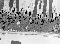
cells of the pigment epithelium behind retina
Bruch'smembrane + choroidea (monkey) |
detail on the left (monkey) |
detail on the right (monkey) |
There are different cells that synthesise pigments and there are also
different kinds of pigments. However, most well known are melanocytes of
the skin with their pigment melanin.
Melanocytes
(Terminologia histologica: Melanocyti) show intensely branched processes
which are free of desmosomes and reach from
the Stratum basale to the
middle of Stratum spinosum (spinous
cell layer) of the skin. By
means of the UV-ray induceable enzyme tyrosinase melanocytes produce brown-black eumelanin
and/or
yellow to red phaeomelanins in
their cytoplasm. A mixture of these pigments
is synthesised in the RER passes the
Golgi-apparatus
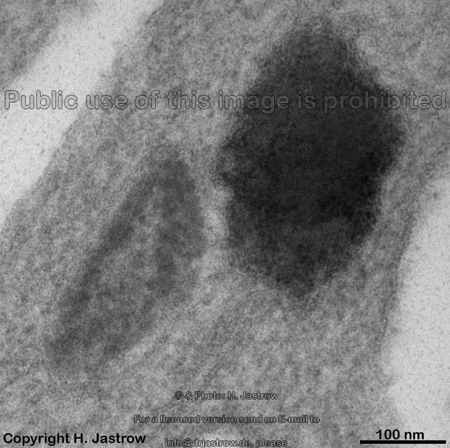
and gets included into vesicles. The latter are called melanosomes
(Terminologia histologica: Melanosomae) and further concentrate pigments
in 4 stages while they get darker and darker. The melanin containing vesicles
(no granules since the are bordered by a membrane) have dimensions of ~100
x 300 nm when cut. They are extruded in toto (i.e. with their membranes)
by the melanocytes and incorporated by neighbouring keratinocytes
(epithelial cells of the skin)
via phagocytosis which then
results in melanin-containing heterophagolysosomes.
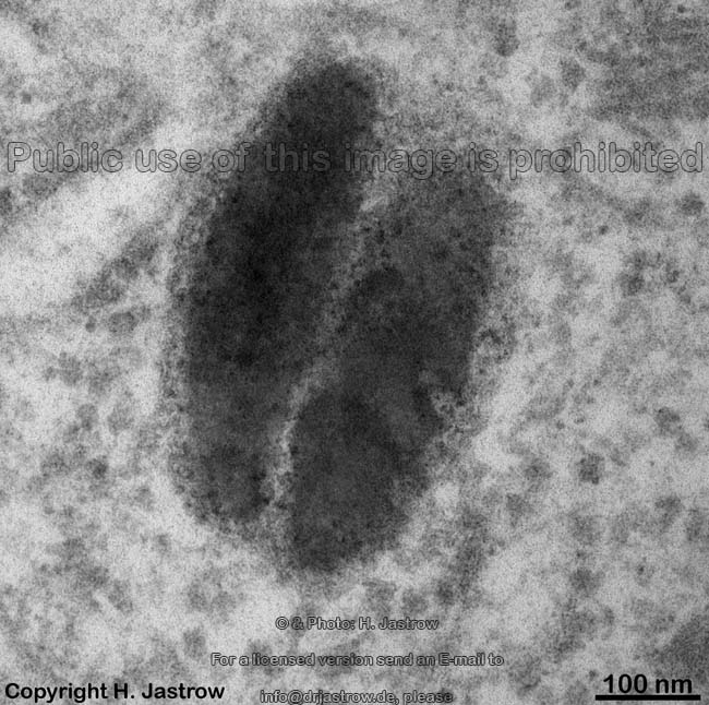
While maturing melanosomes pass the following stages:
1. premelanosome (stage 1 melanosome;
Terminologia histologica: Status 1 Melanosomae, Premelanosomae) have diameters
about 100 x 300 nm and are slightly electron-dense since they derive
from several fused lysosomes that originated
in the Golgi-apparatus. As soon as the homogeneity
of the matrix of premelanosomes shows first condensation the latter become
2. stage 2 melanosome
vesicles (which erroneously often are called premelanosomes; Terminologia
histologica: Status 2 Melanosomae, Vesiculae striatae). The interior of
these special lysosomes shows one or several more electron-dense areas
with ribbon-like aggregated electron-dense granules. The
melanin
proper, however, still is NOT formed since the tyrosinase
still is inactive.
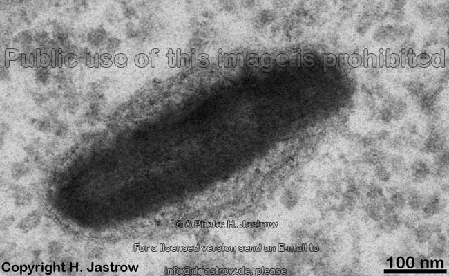
3. stage 3 melanosomes
(Terminologia histologica: status 3 Melanosomae) have
an active tyrosinase and thus now show melanin which gets
attached to matrix filaments. The number of these granules increases and
the areas filled with them grow while the organelles become
4. stage 4 melanosomes
(Terminologia histologica: status 4 Melanosomae) are mature melanosomes.
Sine the matrix here is full of melanin the activity of the
tyrosinase gets more and more reduced. The interior of the mature melanosome
is extremely electron-dense.
The number of melanosomes per keratinocyteis
genetically determined. The paler the skin, the lower their amount.
In Europeans the number varies between 16 132 while in
people originating from countries near the equator it is between 80 160.
An English page with more detailed information and further images is
only available in the professional version
of this atlas.
--> skin, retina,
epithelium,
connective
tissue, eye,
RER,
cytoplasm,
Golgi-apparatus
--> Electron microscopic atlas Overview
--> Homepage of the workshop
Four images were kindly provided by Prof. H. Wartenberg;
other images, page & copyright H. Jastrow.