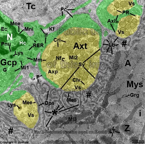
Neuromuscular junction = motor end plate of a skeletal muscle cell from
the tongue (rat)
(for unlabelled original image click here,
please!)

# = subneural junctional fold apparatus formed by deep invaginations
of the cell membrane of the skeletal
muscle cell beneath the free part of
the axon terminal where
transmitter vesicle exocytosis
takes place. A sufficient number of required acetylcholin receptors on
the surface
of the cell membrane of the
skeletal
muscle cell is provided by the considerable increase of cell suface
hereby.
* = invaginations of the cell
membrane of the skeletal muscle
cell, into which the synaptic cleft and the basal
lamina Lb continue;
A = A-band of
the skeletal muscle cell;
Axl
= axolemma (cell membrane of the
axon terminal
Axt);
Axp = Axoplasma (cytoplasm
of the
axon terminal
Axt);
Axt = axon terminal
(terminal boutton, swelling at the end of a motor nerve
fibre);
Clv = Vesiculum clathrinum (clathrin coated endocytotic
vesicle; serves for reuptake of split acetycholin);
Cri = Cristae mitochondriales (inner crests of mitochondria,
formed by invaginations of their inner membrane);
Dps = Densitas postsynaptica (protein rich postsynaptic membrane
thickening located directly beneath the cell
membrane of the skeletal muscle
cell);
Ec = Euchromatin; G
= Golgi-apparatus; Gcp = Gliocytus
periphericus (Schwann's cell); Grg
= Granula glycogeni (beta-glykogen granules);
Hc = Heterochromatin;
Lb
= Lamina basalis (basal lamina);
i
= Stria i (I-band of the
skeletal
muscle cell; contains
actin filaments);
If = intermediate filaments (of the Gcp); Kf =
collagen fibrils of surrounding loose
connective tissue);
Lb = Lamina basalis
(basal lamina); Lys = Lysosoma secundaria
(secondary lysosomes);
Mee = Membrana praesynaptica (pre synaptic cell
membrane = axolemm Axl located above the synapse);
Mes = Membrana postsynaptica (postsynaptic cell
membrane = sacolemm located beneath the synapse);
Mi1 = mitochondria (crista-type
of the Schwann's cell); Mi2
= mitochondrium (crista-type of the axon
terminal);
Mi3 = mitochondria (dark crista-type
of the skeletal muscle cell);
Mm
= Membranae mitochondriales (inner & outer mitochondrial
membranes);
Mne = Membrana nuclearis
externa (outer nuclear membrane); Mni = Membrana
nuclearis interna (inner nuclear membrane);
Mys = Myocytus striatus (striated skeletal
muscle cell);
N = nucleus (of
the Gcp); Nf = neurofilaments
(intermediate filaments of the
axon
terminal
Axt);
P = Plasmalemmata (cell
membranes of the Gcp or skeletal
muscle cell);
Pc = Processus cellulares (immobile processes
of the Gcp);
PR = Polyribosomes (grouped ribosomes);
RER
= rough endoplasmic reticulum (of the
Gcp);
Vs = Vesicula synaptica (synaptic vesicles;
contain the neurotransmitter acetylcholine, which is released by exocytosis);
Sy = 80-100 nm wide synaptic
cleft, that continues into invaginations of the cell
membrane of the skeletal muscle
cell * to form
#;
Tc = Textus connectivus (loose connective
tissue);
Z = Linea Z (Z-line
= Telophragma).
Neuromuscular junctions = motor end plates serve for signal transduction
of motor nerves to skeletal muscle
cells. In other words they are responsible for the innervation of
body muscles. Motor
end plates are chemical synapses releasing
acetylcholine containing neurotransmitter vesicles (Vs) by exocytosis
to stimulate acetylcholin receptors on the postsynaptic cell
membrane of the skeletal muscle
cells. This opens ion channels resultin in an action potential that spreads
rapidly over the whole sarkolemm (cell
membrane of the skeletal muscle
cell) and its deep invaginations into the cell which are called T-tubules
(which are not clearly visible here). The finally resulting contraction
is induced by the ultrafast release of stored calcium ions from
nearby L-tubules (LT = sarcoplasmic
reticulum = smooth endoplasmic reticulum
of the skeletal muscle cell). In contrast
to all other chemical synapses only
the neuromuscular junction shows a special subneural junctional fold
apparatus (#) formed by deep invaginations of the cell
membrane of the skeletal muscle
cell, into which the synaptic cleft and the basal
lamina Lb continue. The acetylcholinesterase, the degrading
enzyme of the neurotransmitter is bound to the collagen 4 network present
in the basal lamina
Lb. The latter further bind the proteoglykan agrin which
induces the aggregation of acetylcholin receptors on the nearby postsynaptic
membrane (Mes). The highest concentration of these receptors is
present on the postsynaptic membrane (Mes) between the secondary
clefts of # which shows a very electron-dense postsynaptic density
(Dps).
--> motor end plate, nerve,
skeletal muscle, synapses
--> Electron microscopic atlas Overview
--> Homepage of the workshop
Labelling by Gregor Wenzel & H. Jastrow, image, page
& copyright H.
Jastrow.


