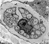
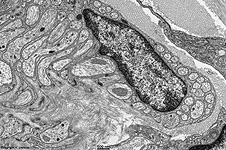
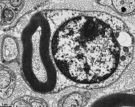
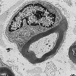
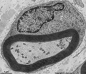
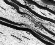
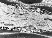
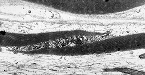
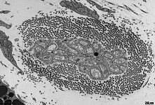
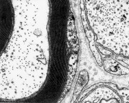
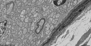
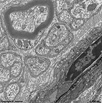
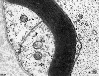
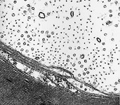
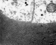
Overview Schwann cells (Gliocyti
peripherici):
Pages with explanations are linked to the
text below the images if available! (Labelling is in German)
Schwann cells (Neurolemmocytes; Terminologia histologica:
Schwannocyti; Neurolemmocyti)
form the myelin sheaths (Terminologia
histologica: Stratum myelini) in peripherical (= nerve tissue apart
from CNS, i.e. apart from brain and spinal cord)
myelinated
nerves and wrap the non-myelinated
nerve cell processes (axons or dendrites) with one layer of their cell
membrane and
cytoplasm. Schwann cells
nutrify
and insulate the axons (impulse conducting
processes of nerve cells to other cells)
as well as dendrites (impulse receiving
processes of nerve cells) of nerves.
They may have a length of
over 100 µm. The
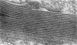
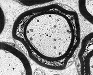
myelin
sheath (Terminologia histologica: Stratum myelini) is formed by
multiple, often far over 20 layers of closely attached cell
membrane of a Schwann cell. The outer
mesaxon (Terminologia histologica: Mesaxon externum) is the connection
of the outer cell membrane to the compact
myelin sheath. The inner
mesaxon (Terminologia histologica: Mesaxon internum) is the connection
between the myelin sheath and the inner part of the cell
membrane of the Schwann cell which is directly opposite the axolemma,
i.e. the cell membrane of the nerve
fibre ensheated by the Schwann cell.
The term terminal Schwann cells
(terminal neurolemmocytes, terminal glial cells; Terminologia histologica:
Schwannocyti terminales, Neurolemmocyti terminales, Cellulae telogliales)
is used for the Schwann cells at the end of nerve cell processes e.g.,
in tactile corpuscules. where
they often form plate like processes in between the dendritic
terminals. Together with
the other Schwann cells and the satellite
glia cells of ganglia they comprise
the peripheral glial cells (Terminologia histologica: Gliocyti peripherici).
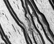
At the border between two neighbouring
Schwann cells a myelin sheath gap called node
of Ranvier (Terminologia histologica: Nodi interruptionis myelini)
is formed along the related nerve fibre. The nodes of Ranvier are the morphological
base for saltatory impulse transduction which is very fast.
Further, there are Schmidt-Lanterman
incisures (Terminologia histologica: Incisura myelini; see this
image), oblique interruptions of the myelin sheath, i.e. myelin clefts
or incisures where some Schwann cell cytoplasm
is present between the myelin layers and gap junctions
are seen in the membrane layers for quick passage of small molecules.
--> classification of nerve
fibres
--> nerve, nerve
tissue, central nervous system
--> Electron microscopic atlas Overview
--> Homepage of the workshop
Ten images were kindly provided by Prof. H. Wartenberg;
other images, page & copyright H. Jastrow.


