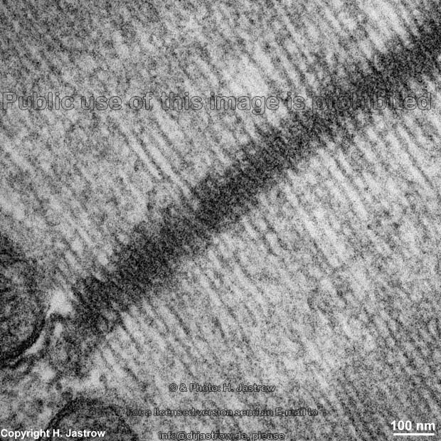
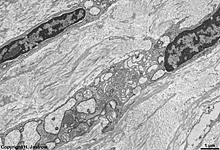
Fibra collagenosa
Abbildungen - pictures
|
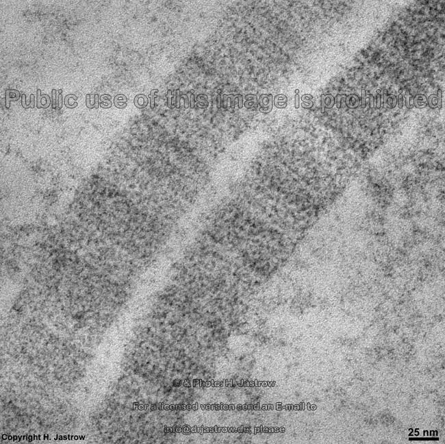
Kollagenfaser; stabile Bindegewebsfaser
aus den in der Abbildung gezeigten Kollagenfibrillen aufgebaut, die aus
einer Tripelhelix mit der Aminosäurefolge Gly-Pro-X bestehend. Es
werden 14 verschiedene Kollagentypen unterschieden, Typ I, II, III, V,
XI zeigen im elektronenmikroskopischen Längsschnitt eine typische
67 nm Periodizität und einen Durchmesser von ~50 nm. Die Synthese
erfolgt aus von Fibroblasten/-cyten gebildeten
Vorstufen, die extrazellulär zum funktionellen Molekül aneinander
gekoppelt werden. Kollagenfasern sind im Allgemeinen sehr zugfest, 5% dehnbar,
anisotrop und meist unverzweigt.
--> weitere Abbildungen und Informationen |
collagenous fibre; a strong fibre consiting of closely associated collagen
fibrils (see image) present in connective tissue,
built of a triple-helix.The sequence of aminoacids of a collagen fibril
is Gly- Pro-X. There are 14 different types, type I, II, III, V and XI
show a periodicy of 67nm when cut lengthwise in electron microscopic sections.
The diameter is about 50 nm. Kollagen precursors are synthecized in fibroblasts/-cytes
and connected in the extracellular space to form the fibres. Fibres are
high-tensile, 5% flexible, anisotropic and usually unbranched.
--> further images and information |
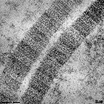
Fibra elastica
Abbildungen - pictures
|
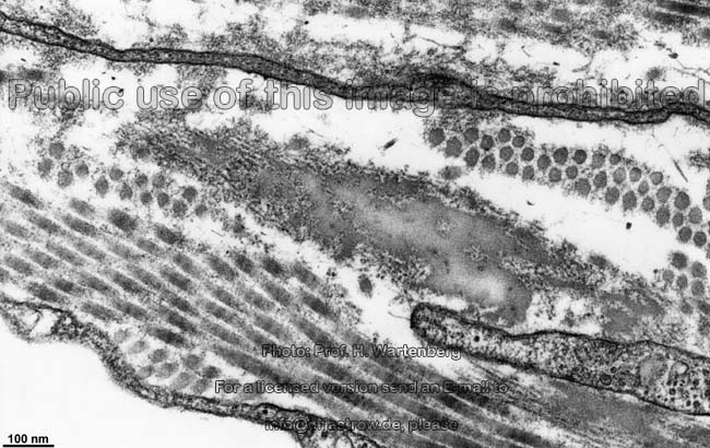
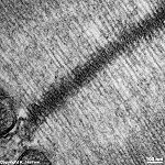
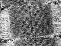
Elastische Faser. Elastische Fasern sind verzweigt und bilden Netzwerke.
Sie bestehen aus 12 nm dicken Mikrofibrillen aus Fibrillin und einem "Kern"
aus Elastin. Das Elastin ist von amorphem Material umgeben, das viel Glycin,
Prolin und etwas Hydroxyprolin enthält.
--> weitere Abbildungen und Informationen |
elastic fibre; these fibres are built around a microfibrillar scaffold
of fibrillin microfibrils (12 nm in diameter). Their centre consists of
elastin. The elastin is surrounded by amorph material rich in glycine,
proline and hydroxyproline.
--> further images and information |
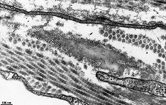
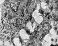
Fibra reticularis
Abbildungen - pictures
|
Retikulinfaser; argyrophile Faser; sehr zarte mit Silber anfärbbare
Fasern im retikulären Bindegewebe. Diese
Fasern bilden feine dreidimensionale Gitter als Grundlage des retikulären
Bindegewebes. Retikuläre Fasern haben Durchmesser von 0,2 1 µm
und sind aus Mikrofibrillen aufgebaut. Dabei handelt es sich um Kollagenfasern
vom Typ III, die insbesondere mit Heparansulfat vernetzt sind und um die
Komponenten, die auch in elastischen Fasern zu
finden sind: 12 nm dicke Mikrofibrillen, Elastin und amorphe Substanz.
--> weitere Informationen |
reticular fibre; argyrophilic fibre, one of the extremely fine silver-staining
fibres found in reticular connective tissue.
These fibres form three dimensional networks in reticular connective tissues.
Reticular fibres have a diameter of 0.2 1µm and consist of a mixture
of different components: collagen type III fibres mostly interconnected
by heparan sulfate, and material also encountered in elastic
fibres: 12 nm microfibrils, elastin and an amorph substance. |
| Fibrillum |
Fibrille (Fäserchen); Fasern mikroskopischer Größenordnung,
die aus kleineren Filamenten besteht; lichtmikroskopisch gerade noch sichtbare
Strukturelemente der Bindegewebsfasern oder
im Zytoplasma von quergestreiften
Muskelzellen; als maskierte Fibrillen in die Kittsubstanz des hyalinen
Knorpels eingebettet. |
Fibril; a small fiber or very small filamentous structure consisting
of smaller filaments, present in striated mucle
cell cytoplasm, masked in haylin
cartilage, present in collagenous fibre bundles. |
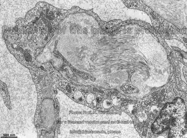
Fibroblastocytus
|
Fibroblast; junge, dem Mesenchym entstammende Zelle mit großem
Zelleib und etwas abgeplatteten Kern; reich
an RER und Golgi-Apparaten;
beteiligt sich an der Bildung von Interzellularsubstanz des Bindegewebes
und wird mit Reduktion dieser Sekretionsfähigkeit zum Fibrozyten.
--> weitere Abbildungen und Informationen |
Fibroblast; a cell deriving from the mesenchyme with large nucleus,
rich in RER and Golgi-apparatuses.
This cell is very active in sectretion of extracellular matrix components
(precursors of collagen, elastin, and reticular protein fibers). When metabolism
decreases the cell transforms into a fibrocyte.
--> further images and information |
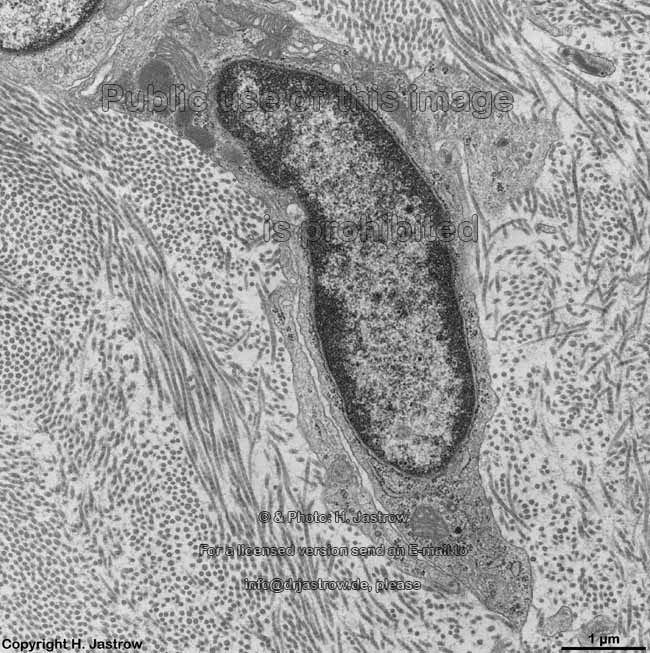
Fibrocytus

|
Fibrozyt; fixe (ortsständige) Bindegewebszelle, länglich,
flächig ausgebreitet, fortsatzreich, mit plattem elliptoidem Kern.
Die Ruheform der Fibroblasten. |
Fibrocyte; an immobile, mature, cell in connective tissue that has
a low metabolism with an elliptois nucleus,
inactive form of a fibroblast |
| Filamenta |
Filamente (Tabelle) |
filaments (table) |
Filamentum actinium
|
Aktinfilament; 2 helikal umeinander gewundene Aktineinzelfäden,
die durch Polymerisation aus freien Aktinmolekülen entstehen; kommen
im Zellskelett sowie gemeinsam mit Myosin
als kontraktile Filamente in der Muskulatur
vor.
--> weitere Abbildungen und Informationen |
actin filament; consists of two helical winding actin molecule chains;
serve as cytoskeletal components or
in muscle contraction. Here actin filaments
are pulled towards the center of the sarcomer by the action of myosin filaments,
and the sarcomere shortens.
--> further images and information |
Filamentum myosinium
|
Myosinfilament, setzt sich aus Myosinmolekülen bevorzugt des Typs
I, II und V zusammen. Diese nur in der quergestreiften
Muskulatur vorkommenden Moleküle weisen einen dickeren Kopfbereich
und einen langen Schwanz auf. Am Übergang, im Halsbereich windet sich
ein Calmodulin Molekül um das Myosin, welches im Typ 1 als Monomer,
in Typ II und V als Dimer vorliegt. Myosinfilamente bestehen aus vielen
rundlich aneinander gelagerten Myosinmolekülen, sind 325 nm lang,
wobei der mittig gelegene Myosinköfchen freie Bereich 160 nm lang
ist. Myosinköpfchen sind 16,5 x 6,5 x 4nm groß. In Gegenwart
von Aktin besitzt Myosin ATPase-Aktivität, bei der Muskelkontraktion
knicken die Myosinköpfchen um 45 Grad ab. In mehreren Schritten ist
eine Verlagerung um 11 15 nm gegenüber den Aktinfilamenten
möglich, wodurch sich die H-Linie und die Sarkomerlänge verringert. |
myosin filament; Myosin filaments are only seen in striated
muscle cells and consist of Myosin molecules, mostly type I,II or V.
The latter have a thicker head (16,5 x 6,5 x 4nm ) and a long tail. In
the neck region between both a calmodulin molecule is winding around the
myosin. Myosin type 1 is a monomer, types II and V are dimers. The myosin
filaments consist of several apposed myosin molecules and have a length
of 325 nm. The middle region, which lacks myosin heads, is 160 nm in length.
When actin is present myosin has an ATPase activity. In the muscle contraction
process myosin heads bend 45 degrees. In several steps a dislocation of
11 15 nm is possible between actin and
myosin filaments, that is the reason for shortening of the H-line and the
sarcomere. |
Filamentum intermediale
|
Intermediärfilament; Filamente, die wegen ihrer Größe
zwischen Mikrofilamenten (8 nanometer; nm) und Mikrotubuli
(25 nm) anzuordnen sind. Sie bilden ein dichtes Netzwerk und sind an Zellkern
und Fleckdesmosomen befestigt.
--> Tabelle der Filamente, weitere
Abbildungen und Informationen |
intermediate filament; smaller than microtubules
(25 nanometers; nm) and thicker than microfilaments (8 nm) they build a
meshwork between the nucleus and spot-desmosomes
--> Table of filaments, further
images and information |
Flagellum
|
Flagelle, Geißel; ein zu kreisender bis schlagender Bewegung
befähigter fadenförmiger Zellfortsatz, der 9x3 + 2 Mikrotubuli
enthält und freien Zellen zur Bewegung dient. Spermien,
Protozoen und einige Bakterien besitzen Geißeln.
--> Abbildungen und weitere Informationen |
Flagellum; a threadlike structure that provides motility for certain
bacteria and protozoa and for spermatozoa.
The circular motion of a flagellum pushes the cells forward. A flagellum
has 9x2 +2 microtubules.
--> further images and information |
-->