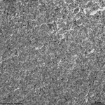
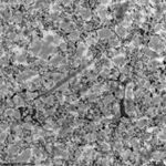
Overview hyaline cartilage (Cartilago
hyalina):
Pages with explanations are linked to the
text below the images if available! (Labelling is in German)
|
|
|
|
|

|
|
|
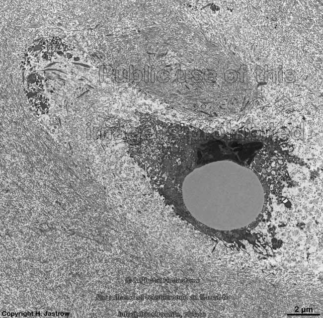
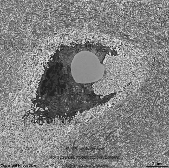
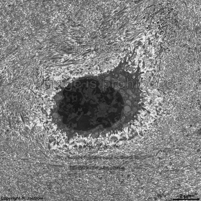
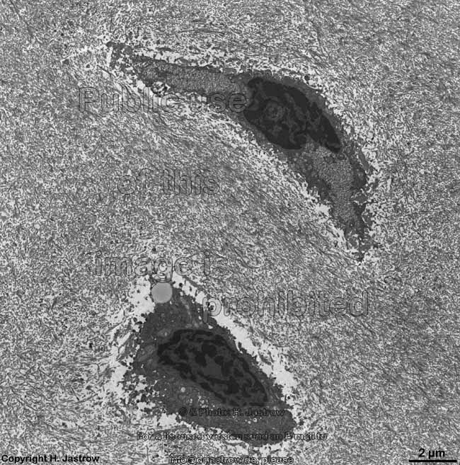
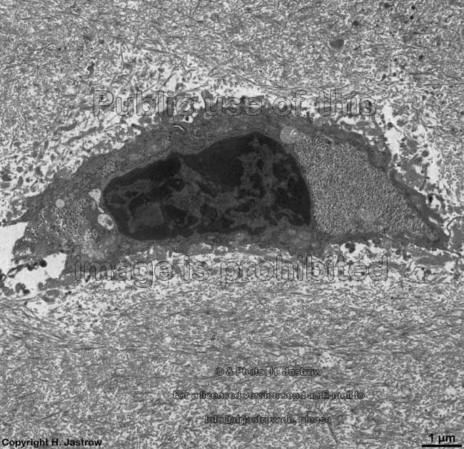
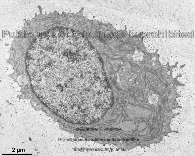
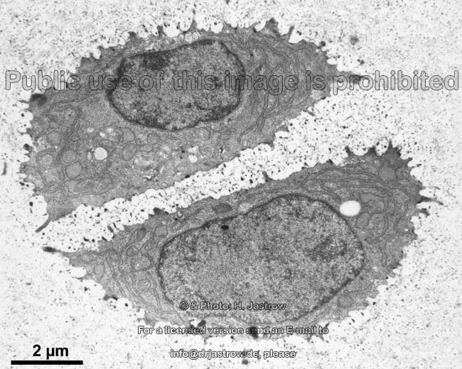
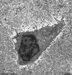
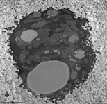
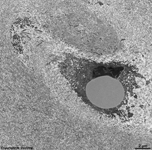
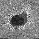
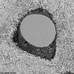
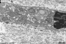
rib cartilage chon-
drocyte 1 (human) |
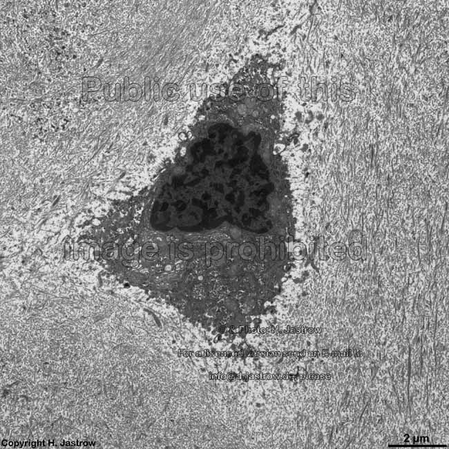
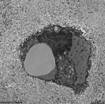
rib cartilage chon-
drocyte 2 (human) |
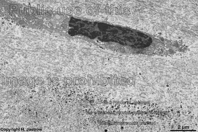
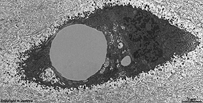
rib cartilage chon-
drocyte 3 (human) |
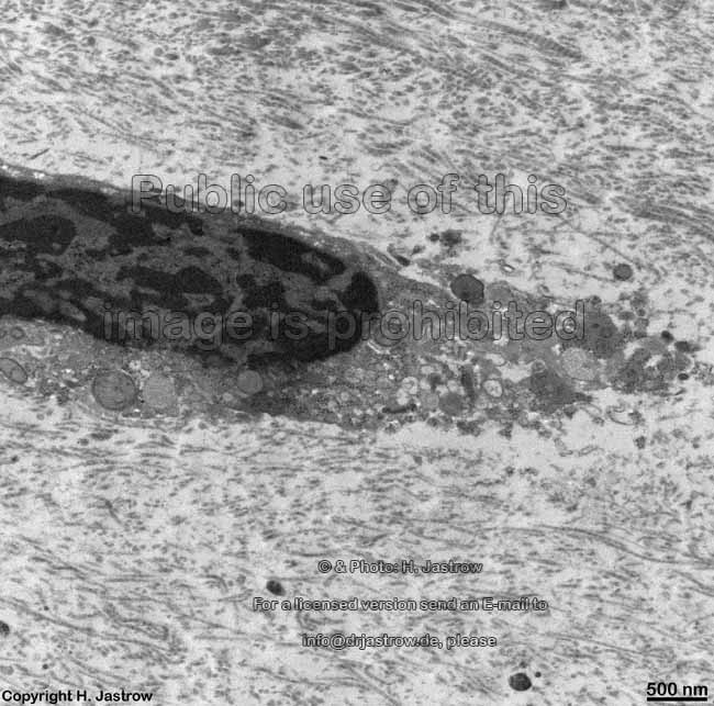
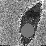
rib cartilage chon-
drocyte 4 (human) |
rib cartilage chon-
drocyte 5 (human) |
rib cartilage chon-
drocyte 6 (human) |
rib cartilage chon-
drocyte 7 (human) |
rib cartilage chon-
drocyte 8 (human) |
|
|
|
|
|
|
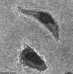
rib cartilage chon-
drocyte 9 (human) |
rib cartilage chon-
drocyte 10 (human) |
rib cartilage chon-
drocyte 11 (human) |
rib cartilage chon-
drocyte 12 (human) |
rib cartilage chon-
drocyte 13 (human) |
rib cartilage chon-
drocyte 14 (human) |
|
|
|

|
|
|
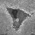
rib cartilage chon-
drocyte 15 (human) |
rib cartilage chon-
drocyte 16 (human) |
rib cartilage chon-
drocyte 17 (human) |
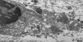
detail thereof
right |
detail in higher magnification |
detail of 17
left |
|
|
|
|

|
|
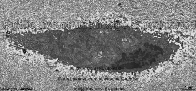
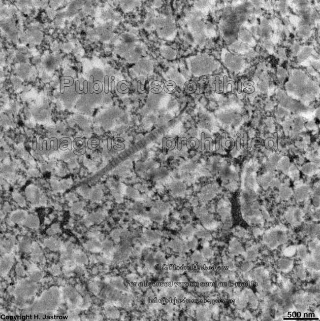
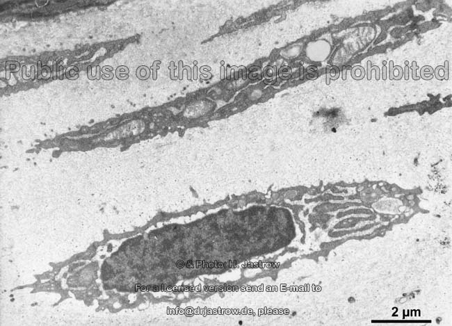
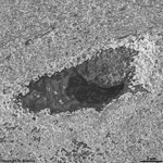
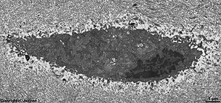
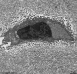
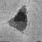
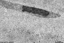
interterritorial substance
rib cartilage (human) |
detail thereof in higher
magnification 1 |
detail thereof in higher
magnification 2 |
detail thereof in higher
magnification 3 |
detail thereof in higher
magnification 4 |
detail thereof in higher
magnification 5 |
The term hyaline cartilage (Terminologia
histologica: Cartilago hyalina) derives from the Greek word hyalos
(glass) and comprises a solid blue-white supporting tissue.
occurrence:
Hyaline cartilage serves as shock absorbing cartilage of joints
in most joints between two bones (diarthroses;
exception: sternoclavicular- and temporomandibular joint) or as stabilising
cartilage.
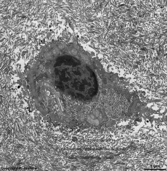
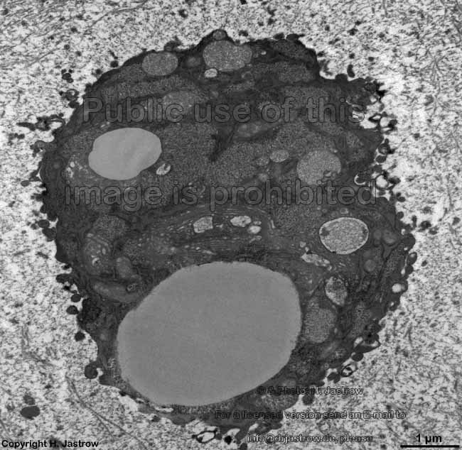
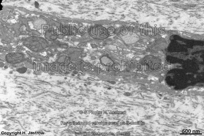
Formation and composition:
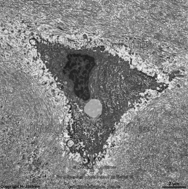
Chondroblasts
(Terminologia histologica: Chondroblasti)
show a little more cell organells
and their metabolic activity is higher than that of chondrocytes
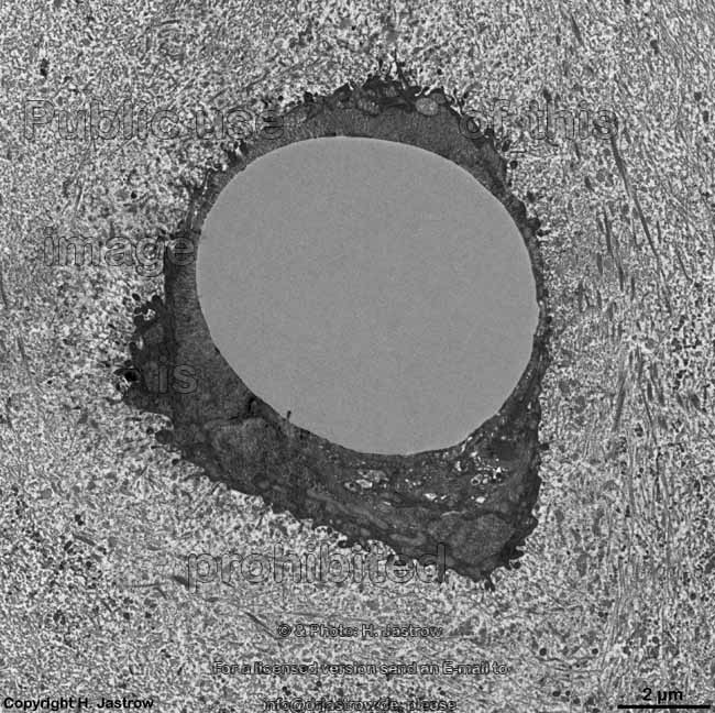
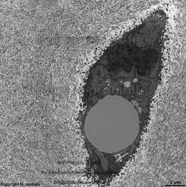
(Terminologia histologica: Chondrocyti) which
comprise the vast majority of cartilage cells in hyaline cartilage
and are in fact just outgrown chondroblasts with reduced activity. The
number of
glycogen particles and few small
lipid
droplets is lower than in chondrocytes. Further, the nuclei
of chondroblasts are larger, about spherical with a considerably higher
content of euchromatin whereas chondrocyte
nuclei are smaller with rather condensed and thus darker chromatin which
predominantly is heterochromatin.
The lipid droplets may fuse in chondrocytes
resulting in larger drops of several µm in diameter. Both cell types
contain a notable amount of rough endoplasmic reticulum
which partly may be dilated by local aggregations of moderately electron
dense proteins, few small Golgi apparatuses
with flattened
dictyosomes giving
raise to tiny
secretory vesicles,
some mitochondria of the crista-type
with a moderate electron-dense
matrix
and few lysosomes and heterolysosomes.
Chondrocytes are ovoid to spherical or even spindle-shaped and contain
many
free ribosomes as well as accumulations
of mainly beta glycogen granules.
The cytoplasm of the cells has a high
electron density. Some of them continue in few, thin and short cellular
processes. Further, vimentin intermediate
filaments contribute to the cytoskeleton.
The slightly basophilic nuclei usually show
several prominent nucleoli. Cellular
metabolism
due to low oxygen content of the surrounding matrix caused by long ways
for diffusion is anaerobic and glycolytic which is responsible for
a high amount of lactic acid secretion into the adjacent matrix.
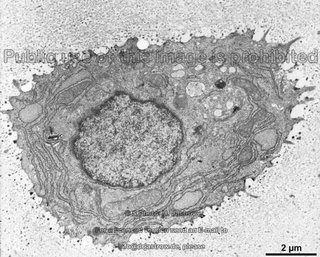
Chondroclasts
(Terminologia histologica: Chondroclasti)
are cells which degrade cartilage. They are present at the borders of hyaline
cartilage during ossification only. These cells are identical to osteoclasts
of bone and odontoclasts which dissolve dentin. They contain several nuclei
and are very large with diameters over 25 µm due to the fact that
they originate from fusing monocytes. Their
cytoplasm is rich in mitochondria of the crista-type
with an electron-dense
matrix and lysosomes
and heterolysosomes. Plenty of cisterns or
RER and larger Golgi apparatuses. Cells secrete hydrochloric acid (HCl)
which solves calcium from mineralized matrix (in bones). Here in cartilage,
however, it is more probable that degrading enzymes which are also secreted
by these cells are the major secretion products of chondroclasts.
In case of mature hyaline cartilage all blood
vessels and nerve fibres
terminate in the perichondrium
(Terminologia histologica: Perichondrium). The
latter consists of an outer tight fibrous layer (Terminologia
histologica: Stratum fibrosum) comprised of woven
connective tissue with some rare fibroblasts
and plenty of elongate thin fibrocytes.
This layer retains tractive power which otherwise would damage the cartilage
proper. Deeper, i.e. directly
attached to the cartilage the chondrogenic layer (Terminologia
histologica: Stratum
chondrogenicum) is able to contribute to formation
of cartilage in young age.
Cartilage matrix (Terminologia
histologica: Matrix cartilaginea)
The intercellular, i.e. extracellular substance around chondrocytes
comprises the cartilage matrix. Due to its high content of aggrecan
(most important proteoglycan with a half-life time of 5 to ~30 days, consists
of ~ 100 chains of chondroitin sulfate + ~30 shorter chains of keratan
sulfate) the cartilage matrix, especially in the cartliage capsule, is
basophilic.
The intercellular substance comprises 70 to 90% of the matrix in hyaline
cartilage of which about 70 to 90 (volume percent;
%) of
the matrix are just water molecules which are bound to the 10
- 30% of organic macromolecules otherwise present.
More details are given in the professional
version of this atlas.
--> elastic cartilage, fibrocartilage,
connective
tissue, resident connective tissue cells,
ground
substance,
elastic-,
collagen
fibres, bone
--> Electron microscopic atlas Overview
--> Homepage of the workshop
Images, page & copyright H. Jastrow.