Overview blood platelets (Thrombocyti):
Pages with explanations are linked to the
text below the images if available! (Labelling is in German)
 |
 |
 |
 |
 |
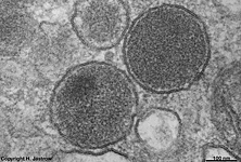 |
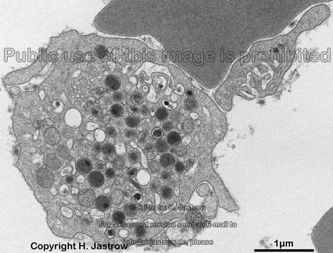
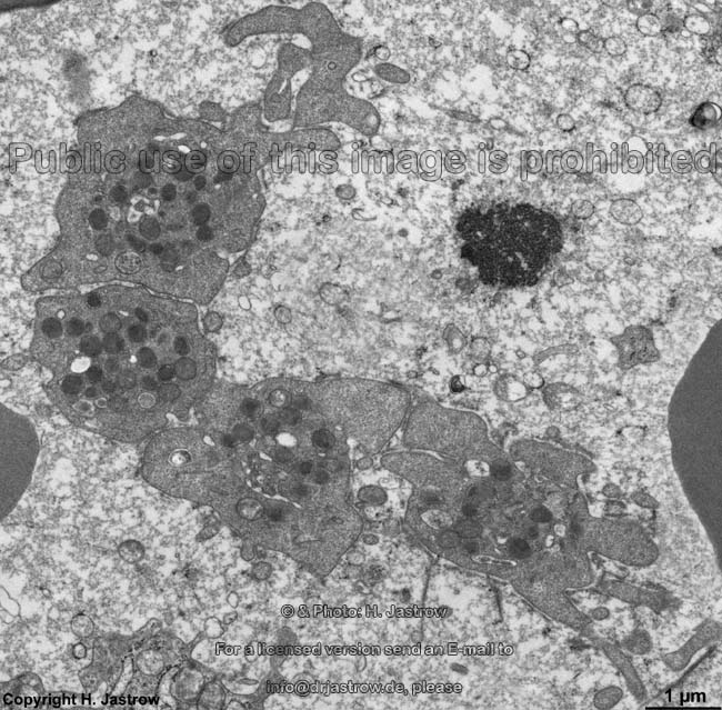
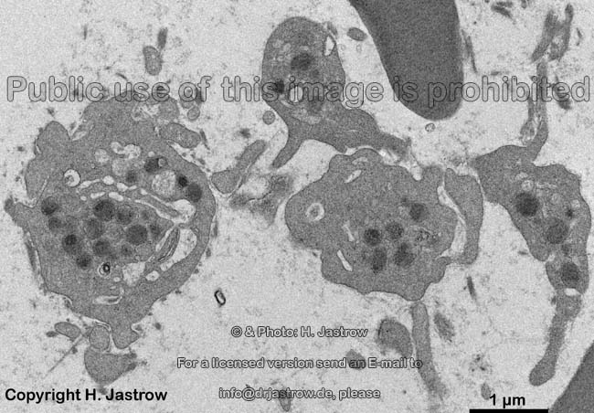
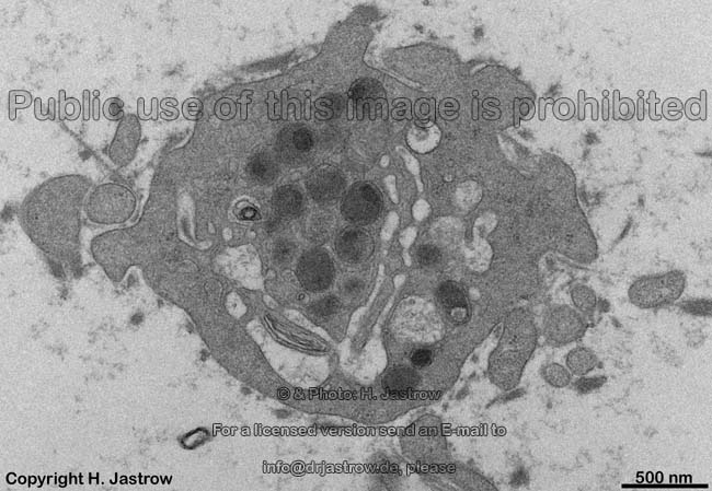
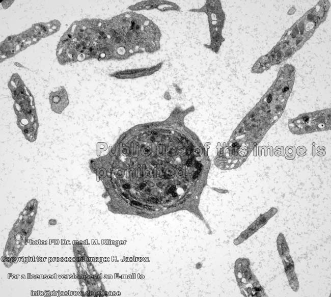
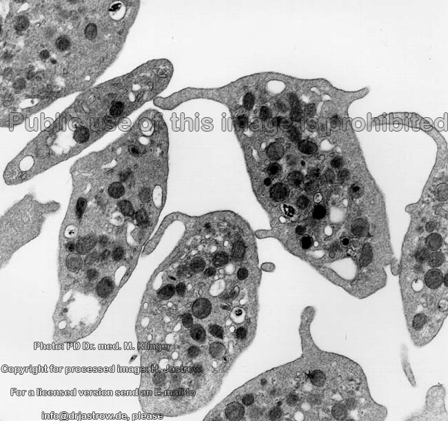
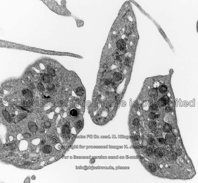
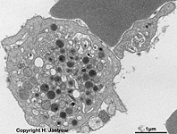
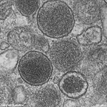
human blood platelets in
different orientations
|
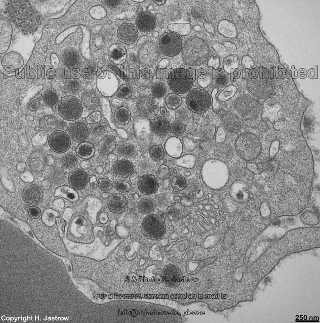
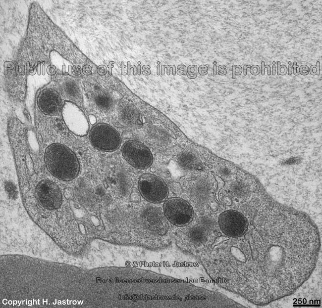
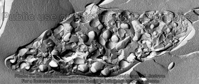
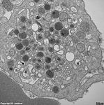
detail 1: cross-sec-
tioned blood platelet |
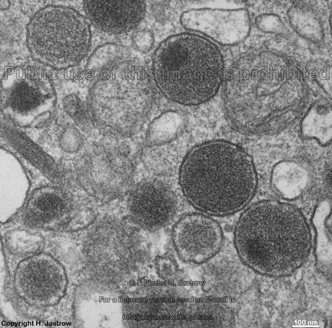
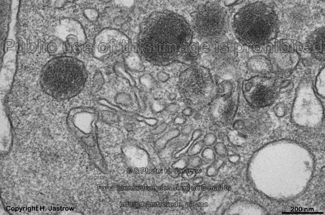
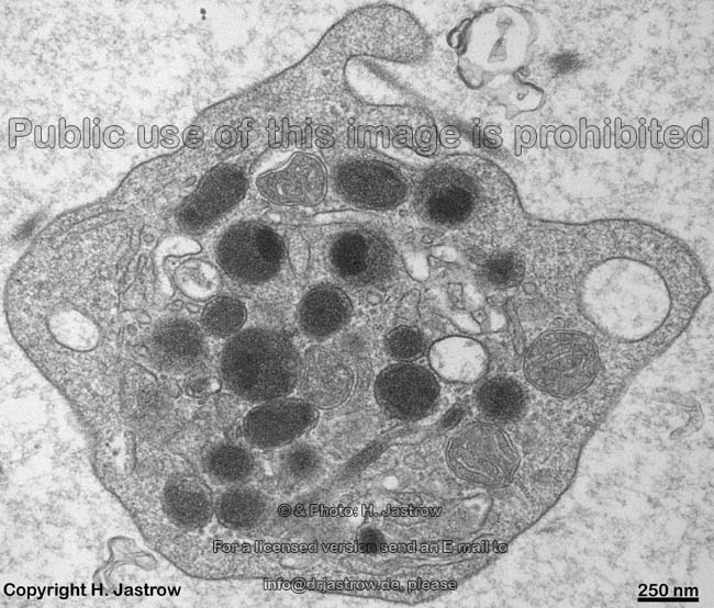
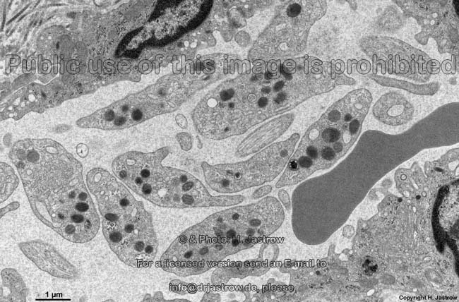
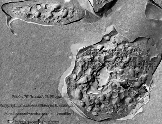
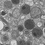
detail 2: cytoplasm
with vesicles |
detail 3: cytoplasm
of one blood platelet |
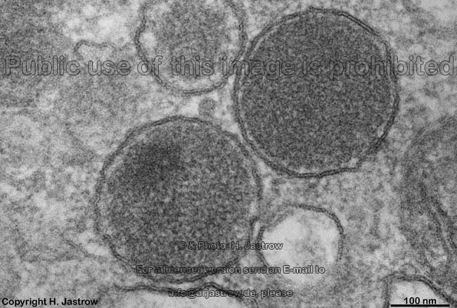
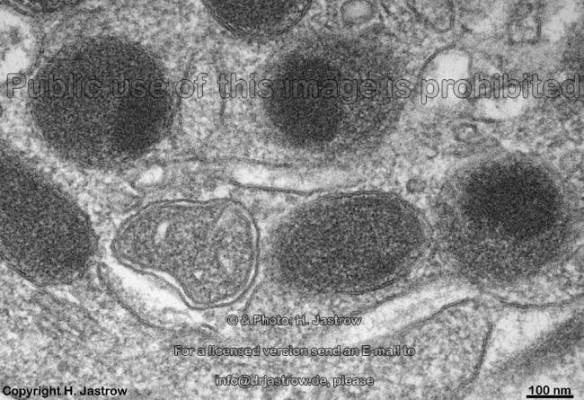
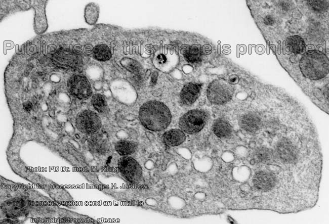
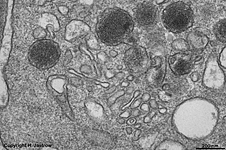
detail 4: different
vesicles |
detail 5: other
vesicles |
Platelets (thrombocytes; Terminologia histologica:
Thrombocyti)
are no complete cells since they lack a nucleus.
The dish-like particles have a diameter of 2 - 4 µm
and aggregate to each other especially in case of a blood coagulation.
In normal human blood about 150,000
- 300,000 thrombocytes are present per mm³. If
the number is less we speak of a thrombocytopenia. The critical value is
about 40,000 since with lower values haemostasis is no longer ensured. The
volume
of platelets is 5.7 - 8.9 femtolitres (µm³). The
thrombocrite
(portion of the platelets related to the entire blood volume) is 13.7
- 26.9%; in children up to 45 % increasing with age. Thrombocytes are
demarked parts of cytoplasm from megakaryocytes
of the
red bone marrow. Their average
life time is 5 - 10 days. They are removed from blood
by phagocytosis performed
by macrophages mainly of liver
(stellate macrophages, i.e. Kupffer
cells) and spleen. Platelets contain free
actin as well as a cytoskeleton
of actin filaments anchored to the
cell
membrane. In this context filamin serves
to bind it to the cell membrane glycoprotein GPIb-IX
and talin for connection to integrin
alpha2b-beta3, the fibrinogen receptor.
Further, the cytoplasm shows mitochondria
of the crista-type and contains myosin, alpha-actinin
and tropomyosin.
Granulomere (Terminologia histologica:
Granulomerus) is the term for the central cytoplasm
area of platelets with plenty of vesicles
which in the light microscope appear to be granules therefore the incorrect
term thrombocytic granules was established in the terminology (Terminologia
histologica: Granula thrombocytica). The granulomere further contains some
beta-glycogen
granules. In Pappenheim's stain this area shows fine blue-purple azurophilic
granules. However, the electron microscope without any doubt reveals membrane-bordered
vesicles.
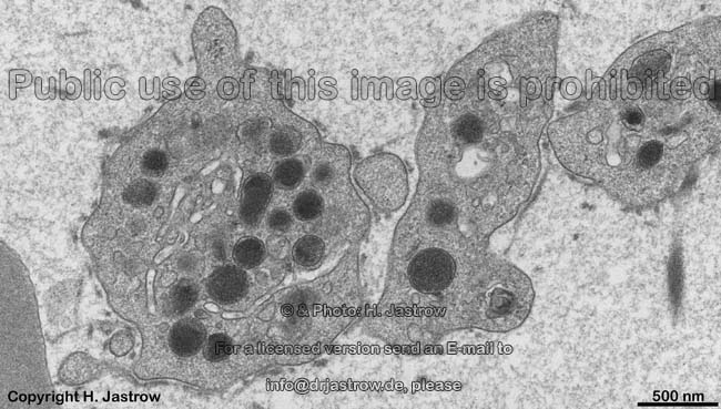
The granulomere contains different kinds of vesicles with diameters about
200 nm which are classified in more or less electron-dense types:
alpha vesicles (Terminologia histologica:
alpha granules; Granula alpha - should be corrected to alpha vesicles;
Vesicula alpha). The less electron-dense alpha-vesicles are an inhomogeneous
population of vesicles corresponding to lysosomes,
peroxisomes
and others which all contain factors for blood cell aggregation e.g., platelet
factor 4 (antiheparin, a heparin binding chemokine), von-Willebrand
factor (vWF), fibronectin, platelet
derived growth factor (PDGF), thrombospondin,
proaccelerin (factor V), factor VIII, beta-thromboglobuline, kallikrein,
alpha2 antiplasmin and fibrinogen. The
electron-dense
vesicles are rare in human platelets, have a small excentrically located
extremely dense core and contain serotonine, adenosin
diphosphat (ADP), histamine,adrenaline, pyrophosphate,
calcium ions (Ca++) and adenosin
triphosphate (ATP). They are classified as
delta-vesicles (Terminologia histologica:
delta granules; dense bodies; Granula deltae - should be corrected to delta
vesicles; Vesicula deltae) which are more common or
lambda-vesicles (Terminologia histologica:
lambda granules; Granula lambdae - should be corrected to lambda vesicles;
Vesicula lambdae).
Hyalomere (Terminologia histologica:
Hyalomerus) is the term for the region bordering the granulomere. The hyalomere
contains slightly blue cytosol
(in Pappenheim's stain) and a marginal microtubular
bundle (Terminologia histologica: Fasciculus microtubularis marginalis)
of 10 - 15 microtubules in circular orientation
which is responsible for the discoid form of platelets. A protein-sugar
layer, the glycocalyx 50 - 150 nm in thickness,
is present on the surface membrane of
platelets. A unique feature
of thrombocytes is the open canalicular system (OCS;
Terminologia histologica: Systema canaliculare apertum) with tubules about
50 nm in diameter consisting of tubules of invaginated cell
membrane reaching deeply into the cell towards the centre of the granulomere.
The tubules are similar to smooth endoplasmic reticulum
but are in fact a cell membrane-bordered
deep invaginations of the extracellular space 50 nm in diameter. Thus a
tiny glycocalyx is present in its lumen. A
second system of tubules with more electron-dense and less wide lumen
termed dense tubular system (Terminologia histologica: Systema tubulare
densum) is branching inside the thrombocytes as well. It corresponds to
the remnants of the smooth endoplasmic reticulum
of the megakaryocyte (from
which the platelet originated). It concentrates calcium ions (Ca++)
and thrombocyte specific peroxidase
as well as enzymes for thromboxan-A2 synthesis
in the lumen and plays an important
role in activation of platelets.
An English page with further detailed information is available in the
professional version of this atlas.
--> Blood barriers, Blood
cells,
Blood vessels
--> Electron microscopic atlas Overview
--> Homepage of the workshop
Some images were kindly provided by PD Dr. Klinger;
other images, page & copyright H. Jastrow.