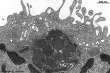
Overview liver (Hepar):
Pages with explanations are linked to the
text below the images if available! (Labelling is in German)
 |
 |
 |
 |
 |
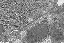 |
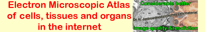
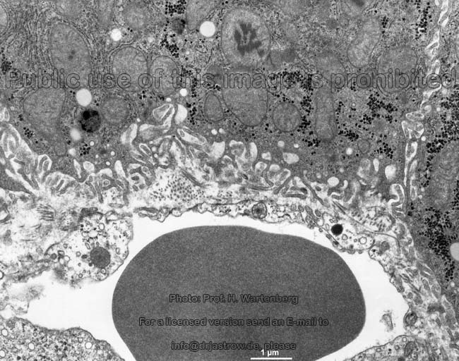
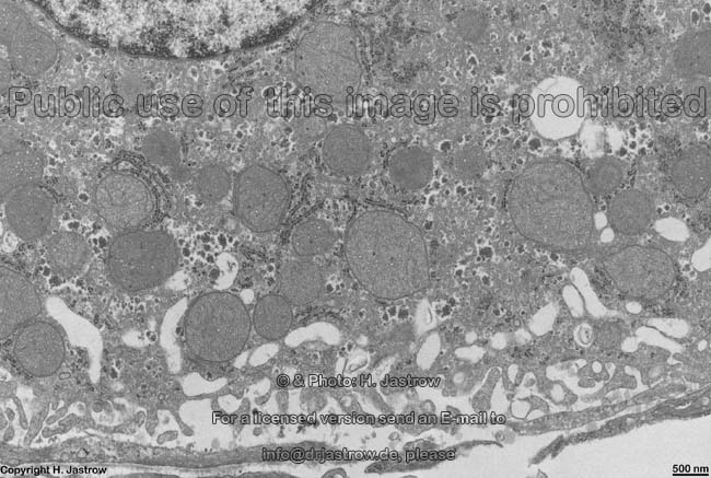
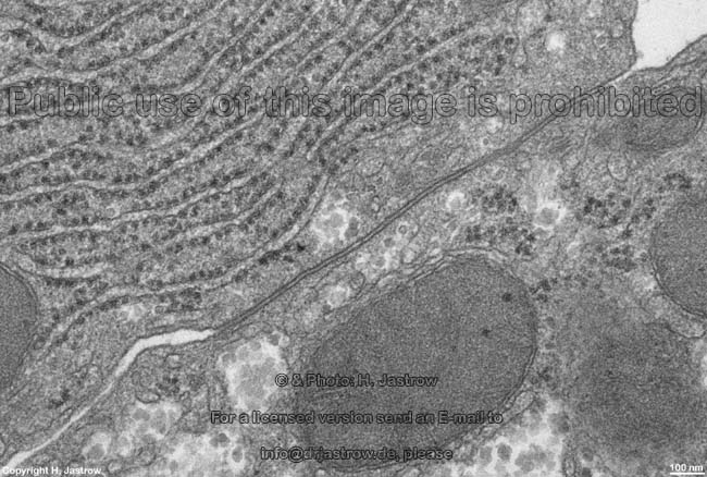
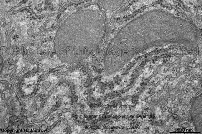
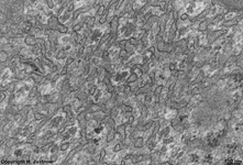
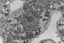
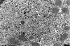
Disse's space, apical
cytoplasm (rat) |
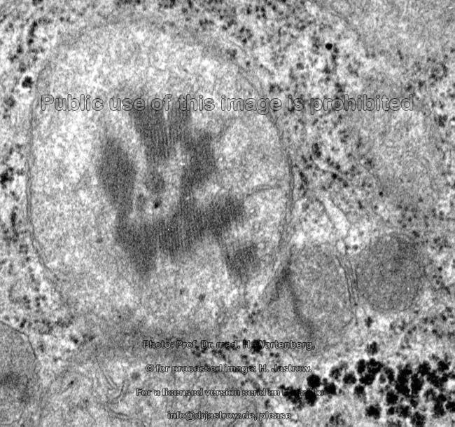
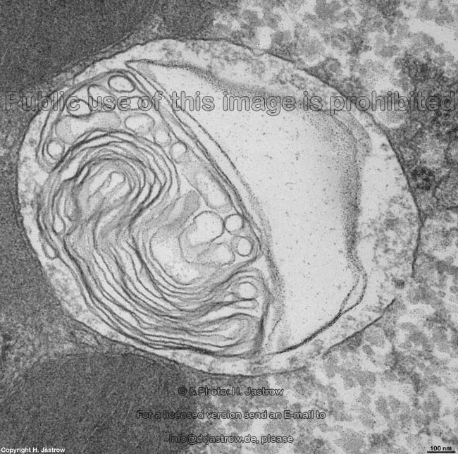
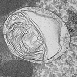
cytoplasm: multilam-
mellar body (rat) |
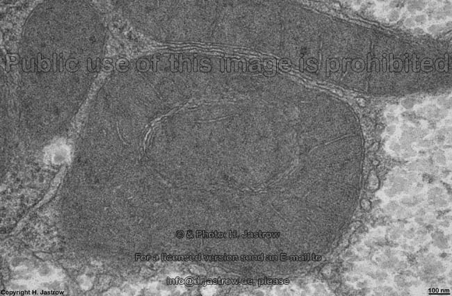
cytoplasm: dark Mito-
chondria of crista type (rat) |
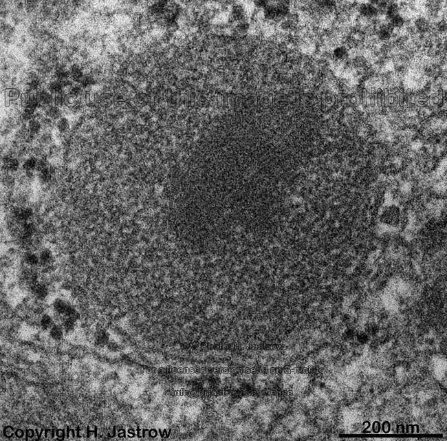
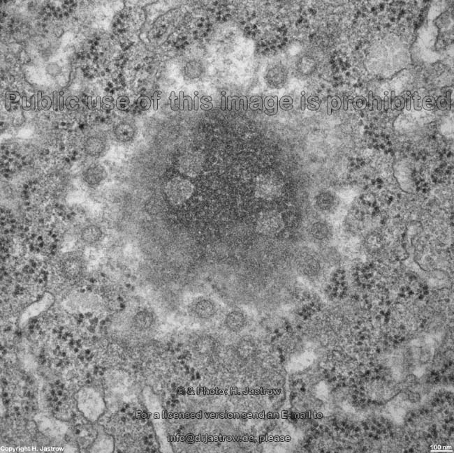
cytoplasm: peroxysome
(rat) |
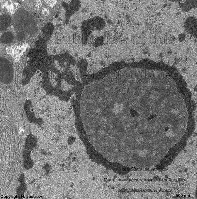
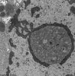
nuclear pores of a
hepatocyte (rat) |
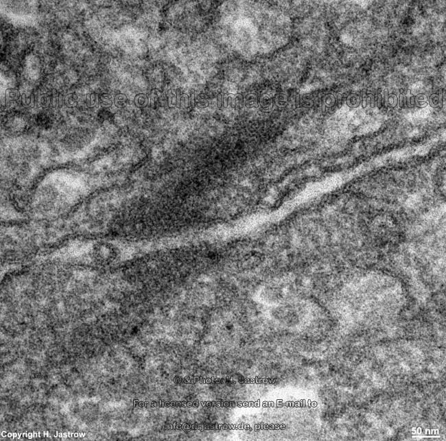
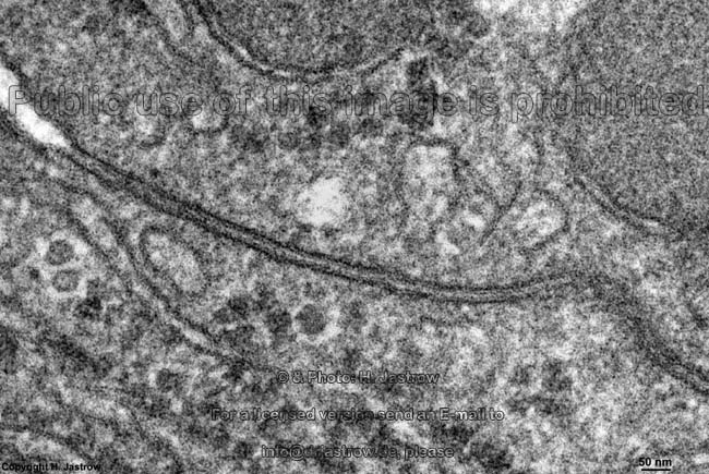
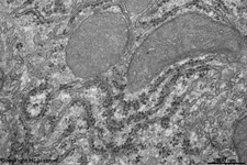
Zonula occludens in between
2 hepatocytes (rat) |
 |
 |
 |
 |
|
|
 |
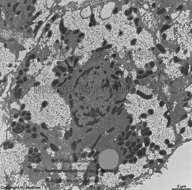
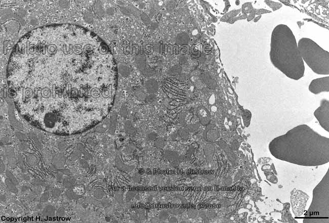
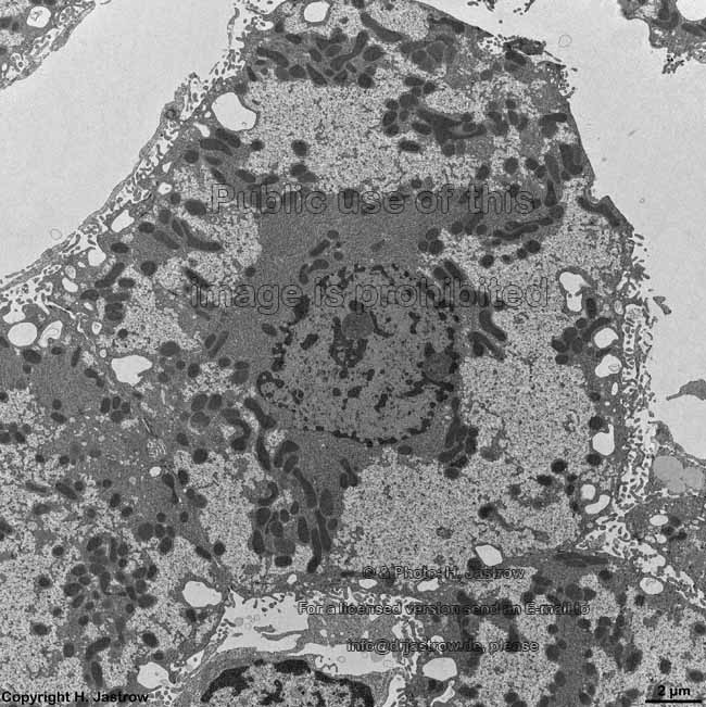
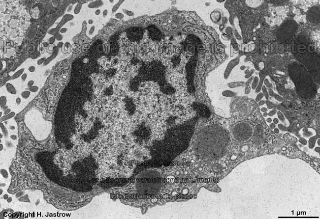
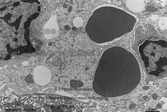
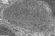
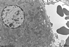
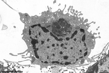
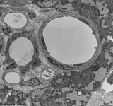
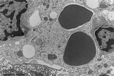
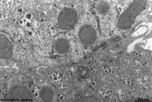
| hepatocyte 2 (rat) |
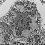
ITO cell 2 (rat) |
ITO cell 3 (rat) |
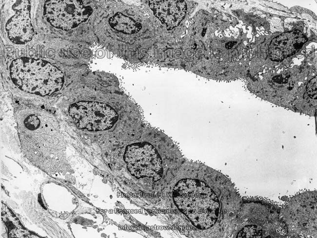
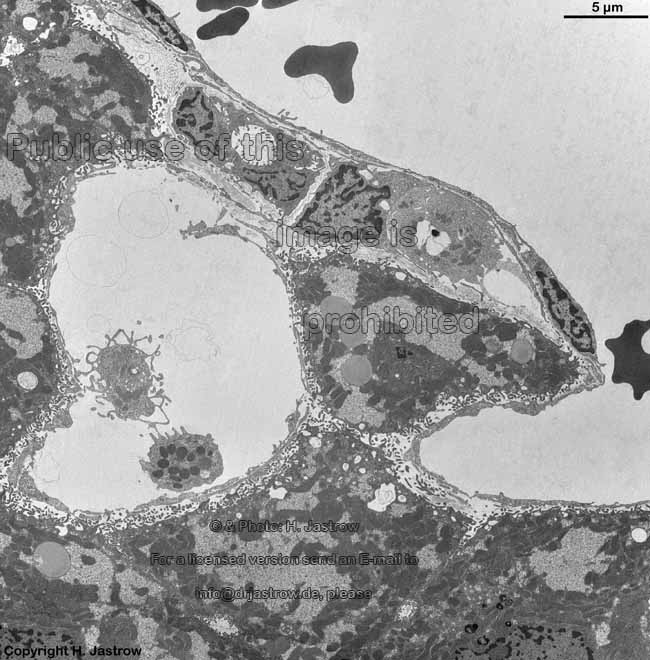
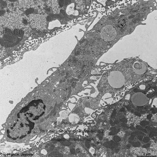
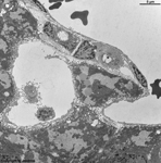
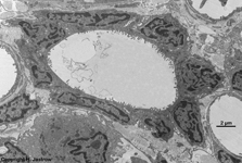
fenestrated sins endo-
thelial cell 1 (rat) |
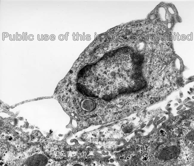
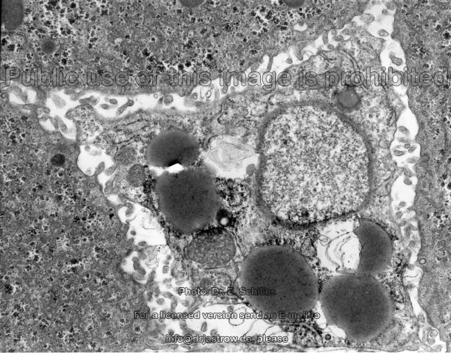
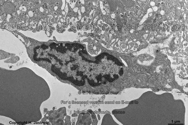
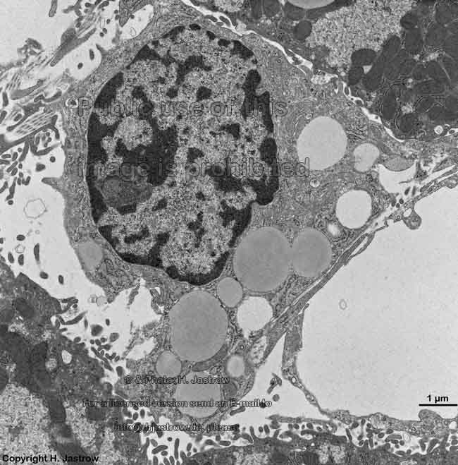
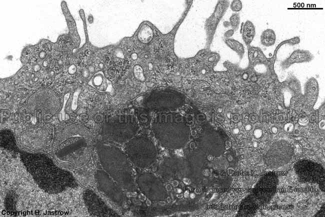
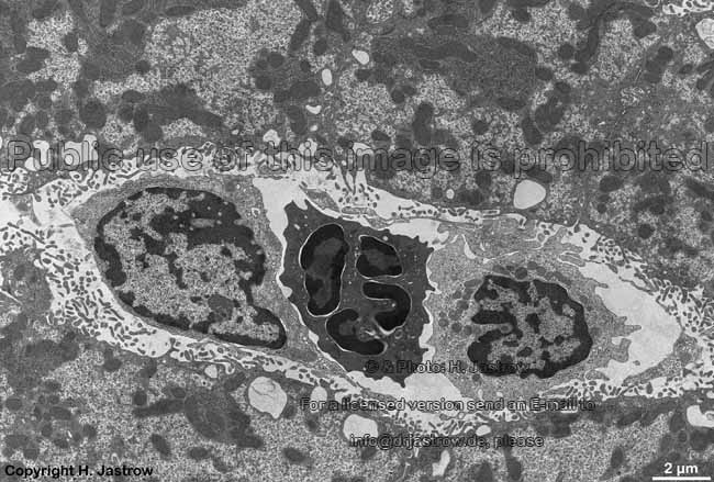
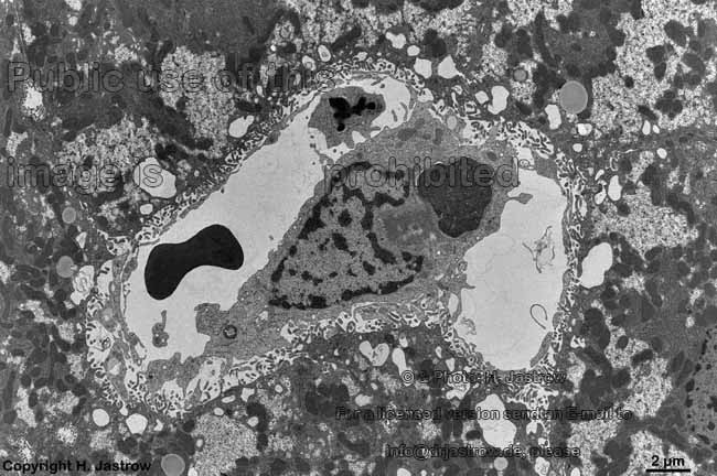
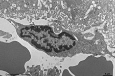
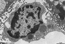
Kupffer cell 2
(rat)* |
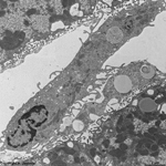
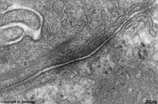
Herring canal,
endothelial cells (rat)* |
hepatocyte 3 (rat)* |
 |
 |
 |
|
|
|

|
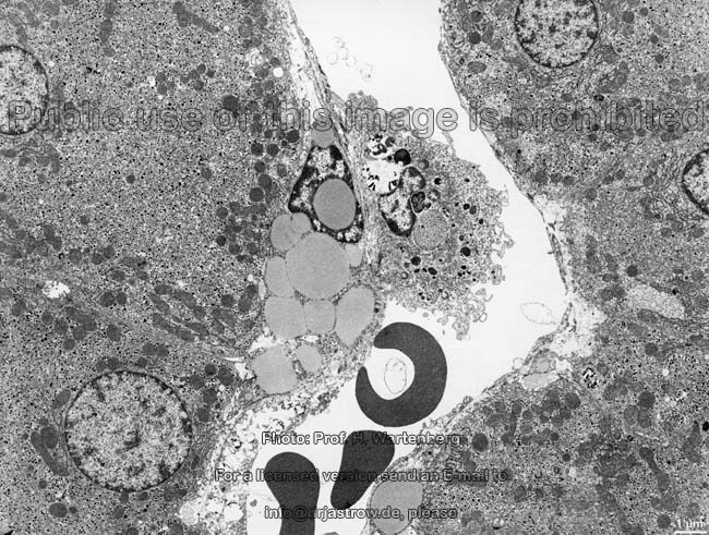
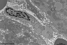
| ITO cell 4 (rat) |
fenestrated endothelial
cell of a sinusoid (rat) |
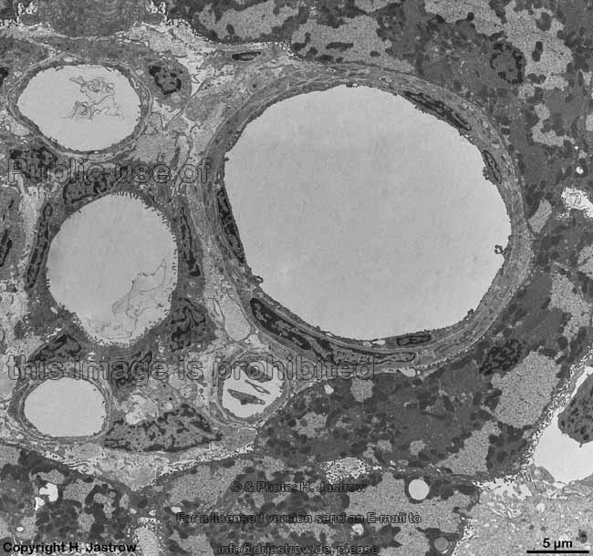
bile duct (rat) |
Glisson's triad: arteriole
+ bile duct (rat)* |
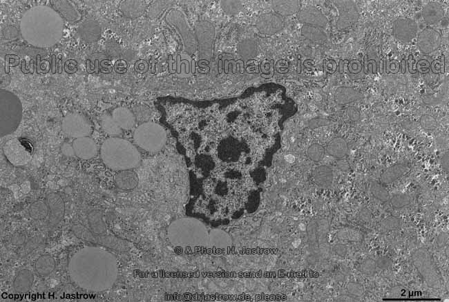
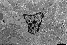
Kupffer cell 3
(rat)* |
detail of Kupffer cell 2:
large phagolysosome |
granulocyte, Kupffer cell +
endothelial cell (rat)* |
 |
|
|
 |
 |
|
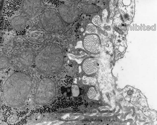
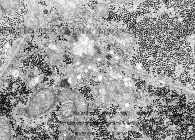
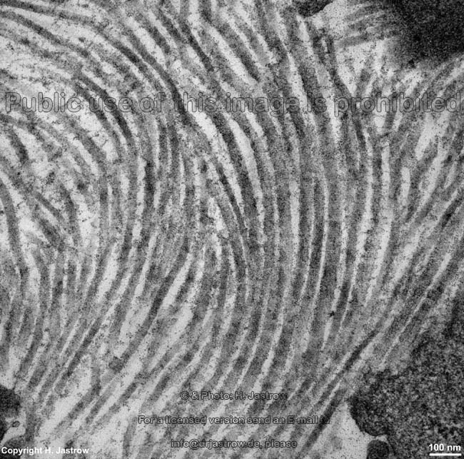
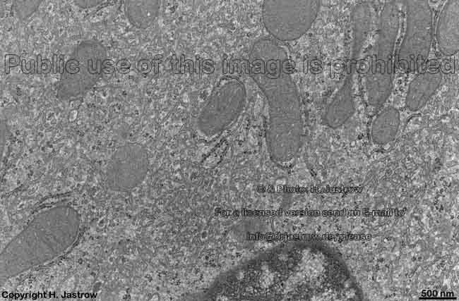
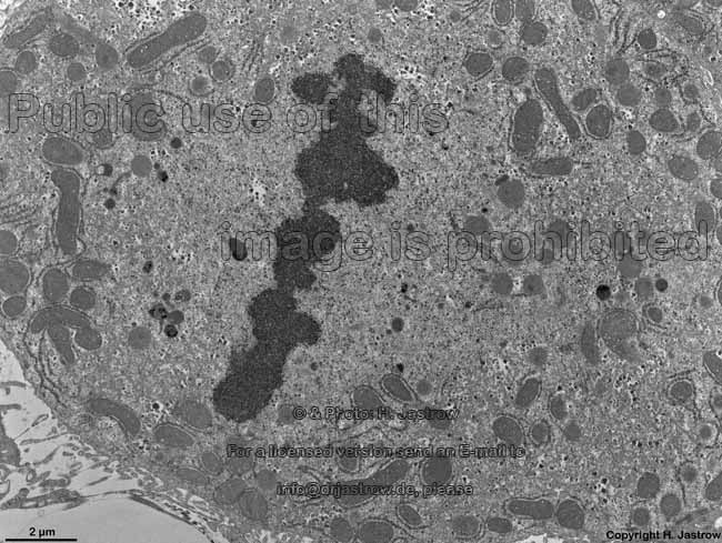
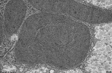
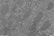
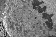
hepatocyte 3 with excellently
preserved cytoplasm (rat) |
detail 1: cytoplasm with
RER, SER + mitochondria |
detail 2: other cyto-
plasmatic region |
detail 3: smooth endo-
plasmic reticulum |
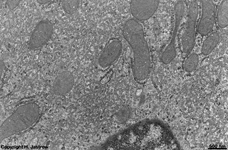
detail 4: mitochondria
and RER |
detail 5: dark mito-
chondrium of crista-type |
 |
 |
 |
|
|

|

|
| ITO cell 5 (rat) |
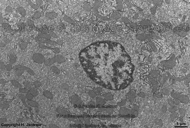
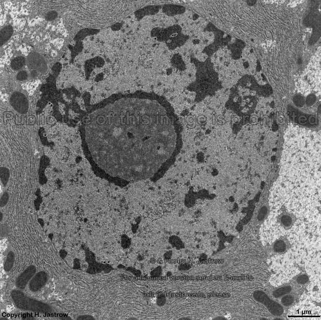
hepatocyte 4 (rat)* |
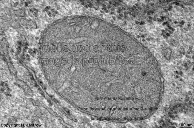
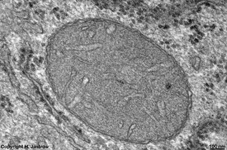
detail 1: nucleus |
detail 2: nucleolus |
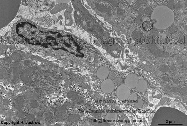
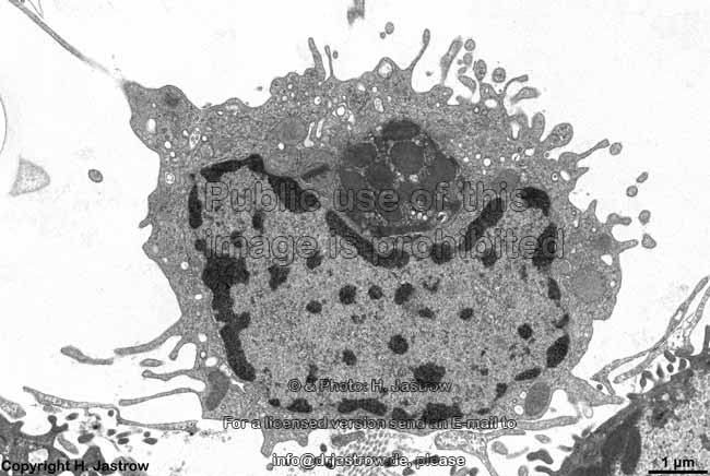
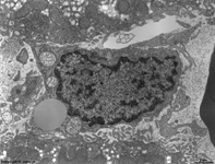
Kupffer cell 4 with
phagocyted erythrocytes (rat) |
Kupffer cell 5 with large
phagolysosome (rat)* |
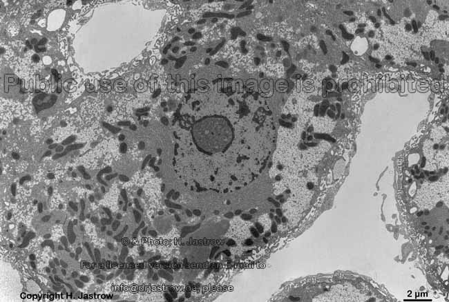
Pit cell = liver specific
T-lymphocyte (rat)* |

|
|
|
|
|
|
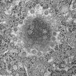
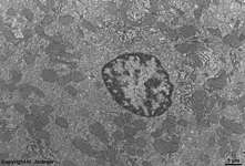
mitosis of a
hepatocyte (rat) |
detail 1 thereof |
detail 2 with spindle fibres
(microtubuli) + organells |
cell-to-cell contacts between
hepatocytes (rat) |
Zonula adhaerens between
2 hepatocytes (rat) |
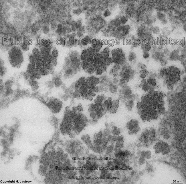
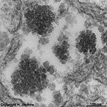
alpha-glycogen-
granules (rat) |
Liver lobule classifications:
The functional tissue of the liver,
i.e. the liver parenchyme is divided in hepatical
lobules (Terminologia histologica: Lobuli hepatici). From different
points of view the following classifications have been established:
1. The hepatocytes are ordered in polygonal
or classical hepatical lobules (Terminologia histologica:
Lobuli hepatici classici; Lobuli hepatici polygonales) which show the central
veins in their centres. These veins drain the blood
into the branches of the liver veins. The
hepatocytes
are arranged in 1 - 1.5 millions polygonal, ~2 mm³ huge
classical lobules in the centres of which the central
veins are located in this classification. From here the hepatocytes,
which are arranged in dense, parallel laminas, run like spokes of a wheel
in direction of the portal fields. Regarded
three-dimensionally they form six-edged columns resembling honeycombs.
The 0.5 - 0.6 mm long sinusoids
run in between the hepatocyte plates and slowly
carry blood from the periphery to the central
vein into which they drain. The hepatocyte laminas are bi-layered with
bile
canaliculi running at their cell membrane
borders. These canaliculi start close to the central
veins and lead the bile fluid
from here towards the bile ductules in the
periphery. Thus the bile flow is in
opposite
direction to blood from the centre
of the lobule outwards. Classical hepatical lobules have a height
of about 2 mm and their average diameter is in between of
1
- 1.3 mm. The
portal
areas (portal canals, portal zones; Terminologia histologica:
Spatia portalia) are located at the border between three or more lobules.
They consist of loose connective tissue
and contain interlobular capillaries,
a small lymphatic vessel as well as the portal
triad (Glisson's triad; Terminologia histologica: Trias
hepatica). The latter is comprised of the following closely apposed vessels:
an interlobular artery (brings oxygen-rich
blood from Arteria hepatica propria),
an interlobular vein (carries blood from
the portal vein) and a small bile
duct (Ductus bilifer interlobularis; carrying bile
fluid from bile ductules to the larger
bile ducts finally the hepatic duct).
2. liver acinus (Terminologia
histologica: Acinus hepaticus): in this classification tissue oxygen
content and thus the terminal arteriols
are of main interest.
3. portal lobules (Terminologia
histologica: Lobuli portales). In this classification the portal
canals with interlobular vein, arteries
and bile ducts are regarded as centres
of the lobules. The 3 closest central veins
then form the border lines of the rather triangular lobules which
in three dimensions form tri-edged columns.
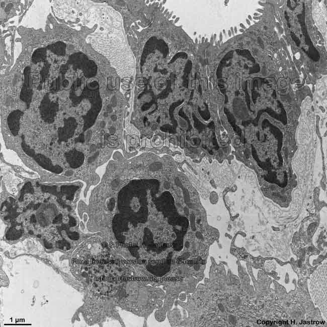
Cell types of the liver and their functions:
A. hepatocytes, liver parenchyma
cells or liver epithelial cells (Terminologia histologica: Hepatocyti)
comprise the parenchyma (functional tissue) of the liver as the most important
kind of cells in point of view of function. They are also called liver
epithelial cells and have diameters of 20 - 30 µm and
a polyhedral shape with 6 or
more relatively straight edges in cross-section. Hepatocyte
laminas, i.e. hepatocytic plates or hepatic trabeculae (Terminologia
histologica: Laminae hepatocyticae) consist of larger arrays of directly
attached hepatocytes and in the three dimensions
form the hepatic muralium (Terminologia
histologica: Muralium hepaticum) by attachment onto each other resulting
in a network of cells of about parallel standing plates in between which
the sinusoids branch. About 20 - 25% of
the hepatocytes, due to their enormous metabolic activity show
2 nuclei.
Such cells are more often encountered in the periportal
zone I. About 50 - 60% show nuclear diameters
of ~ 15 µm and are tetraploid (4-fold chromosome
set) and the largest nuclei with diameters of ~20 µm are even octaploid
(8-fold chromosome set) while the remaining
hepatocytes have a a diploid chromosome
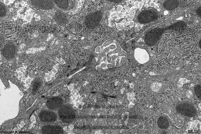
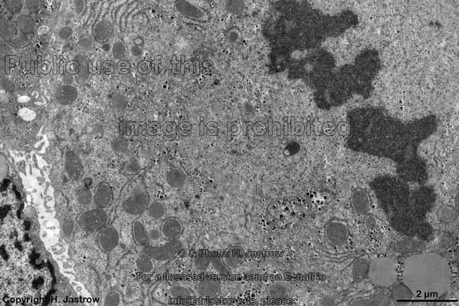
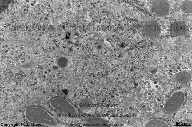
set. Hepatocyte cytoplasm
usually is rich in
alpha-glycogen granules,
shows many dark mitochondria of the crista-type
and plenty of rough (RER)- and smooth
endoplasmic reticulum (SER). In the periportal
zone amount of RER is much higher than that
of SER whereas close to the central
veins both are about equal. Liver parenchyma cells contain about 500
peroxisomes
each with diameters of 200 - 800 nm which are involved in cholesterin-
and bile acid synthesis.
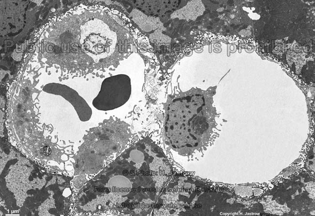
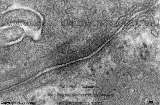
B. perforated endothelial
cells (Terminologia histologica: Endotheliocyti perforati): The
flat
endothelial
cells of the liver sinusoids form a discontinuous
endothelium with plenty, partly over 100 nm wide, gaps lacking
a basal lamina. Neighbouring
endothelial
cells overlap, but do NOT form tight
junctions in between each other. Further, the cells show some hundreds
of open transcellular pores about 100 nm in diameter
which often are closely adjacent forming cribriform plates.
The stellate macrophages
also known as
Kupffer cells (Terminologia histologica: Macrophagocyti
stellati) are large
macrophages located
in the lumen of sinusoids which keep their position in the streaming
blood
by several pseudopods which they stretch
through endothelial pores into
Disse's space. With their other free pseudopods they reach into the lumen
trying to catch erythrocytes and other larger slow moving particles or
cells.
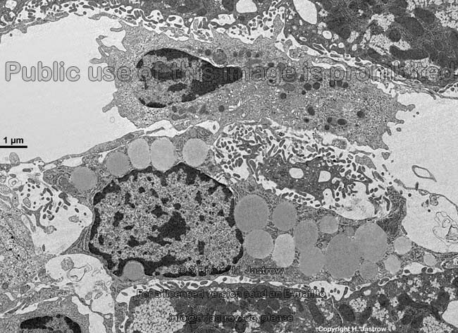
D. ITO-cells which are also called
perisinusoidal cells, fat storing cells or hepatic stellate cells (Terminologia
histologica: Cellulae perisinusoidales; Cellulae accumulantes adipem)
store fat and vitamin A. They are located in the perisinusoidal
space (Disse's space).
E. Pit cells (hepatic natural
killer cells or hepatic NK cells; Terminologia histologica: Cellulae necatoriae
hepaticae) which are specialised T-lymphocytes
serving for immune defence processes are also located in Disse's
space.
F. Fibrocytes
(Terminologia histologica: Fibrocyti) and fibroblasts
(Terminologia histologica: Fibroblasti) which have a higher metabolic activity
are usually only present in the loose
connective tissue around the classical hepatic
lobules which in humans in contrast to pigs only in case of a liver
cirrhosis completely surrounds the lobules but is restricted to the
portal
and periportal fields under normal conditions.
Only close to the central viens few
fibrocytes
are commonly seen.
G. Cholangiocytes
(Terminologia histologica: Cholangiocyti) comprise the isoprismatic
epithelial cells of bile ducts.
H. Oval
cells (Terminologia histologica: Cellulae ovales) are the cuboidal
epithelial cells with a characteristic single oval nucleus
which comprise the epithelium of Hering canals
with low differentiation which are regarded as stem
cells from which cholangiocytes as well as even hepatocytes shall differentiate.
--> bile canaliculi, bile
ducts and gall bladder
--> Electron microscopic atlas Overview
--> Homepage of the workshop
Some images were kindly provided by Prof. H. Wartenberg
or Dr. E. Schiller; other images, page & copyright H. Jastrow.