Overview neutrophil granulocytes
(Granulocyti
neutrophili):
Pages with explanations are linked to the
text below the images if available! (Labelling is in German)
|
|
|
|
|
|
|
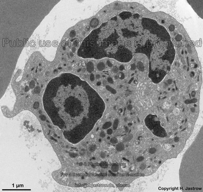
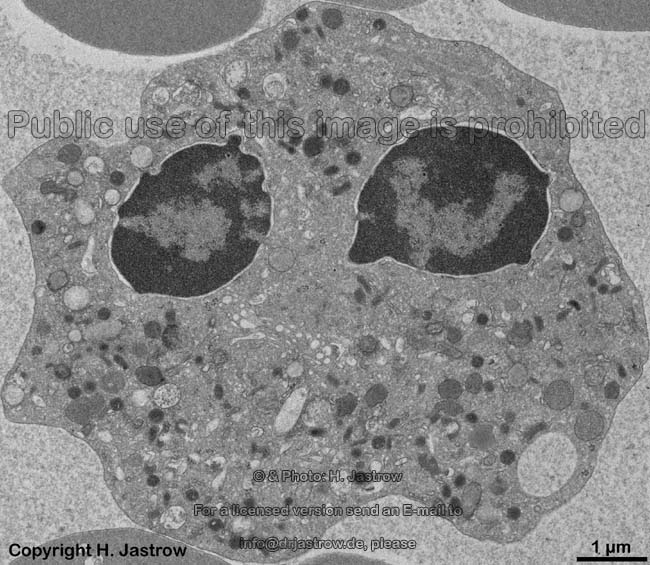
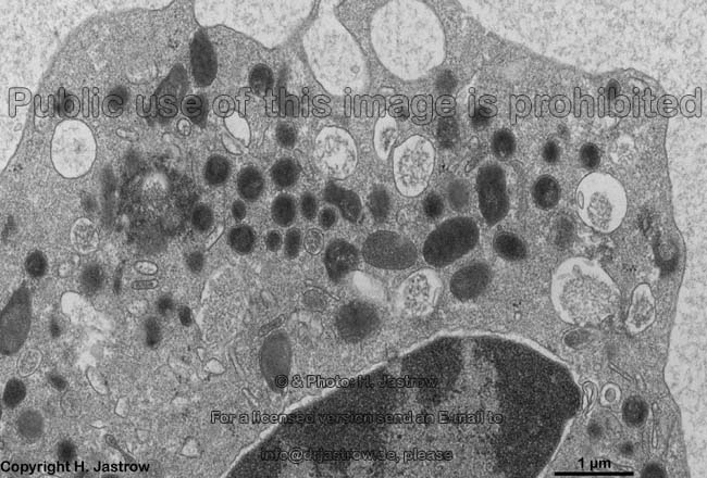
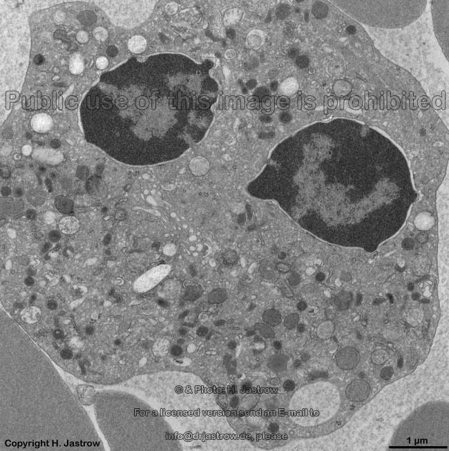
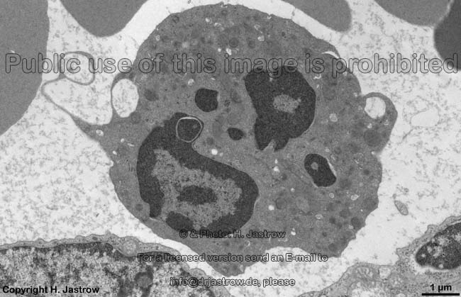
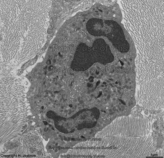
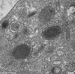
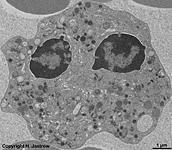
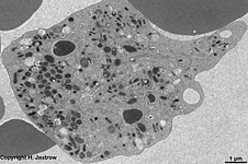
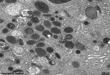
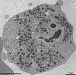
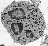
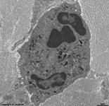
human segmented
neutrophil granulocyte
|
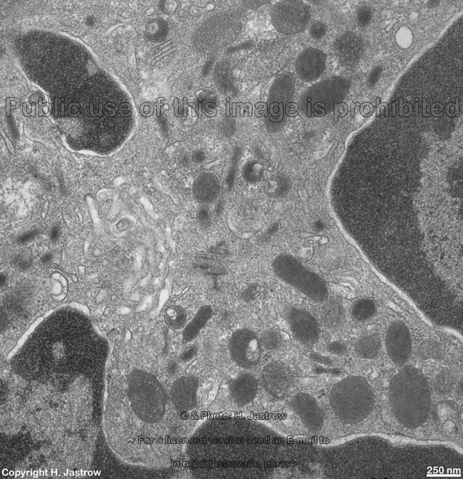
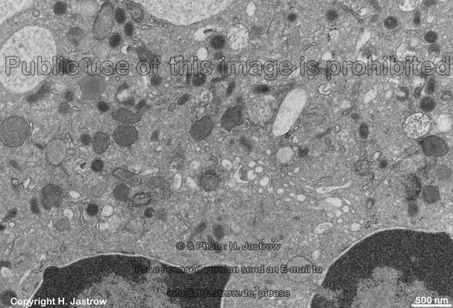
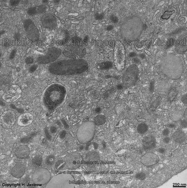
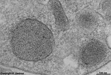
detail 1:
centarl cytoplasm |
detail 2:
cytoplasm with vesicles |
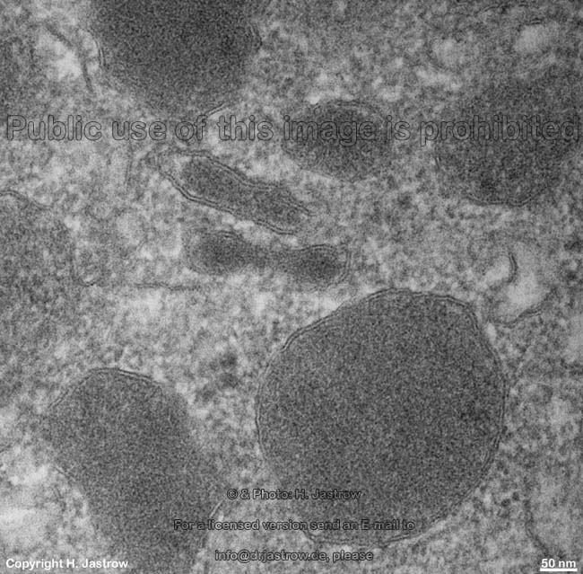
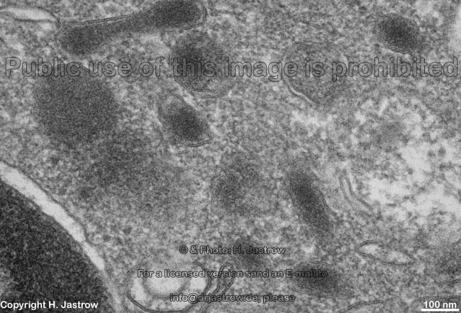
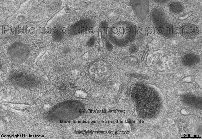
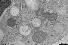
detail 3:
different vesicles |
detail 4:
other vesicles |
detail 5:
some more vesicles |
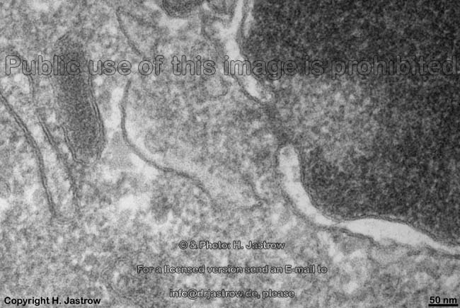
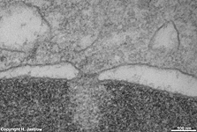
Detail 6: nuclear pore +
specific vesicle |
|
|
|
|
|
|
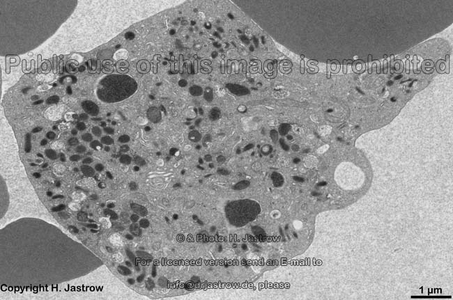
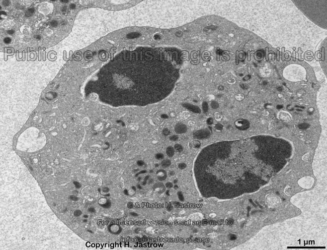
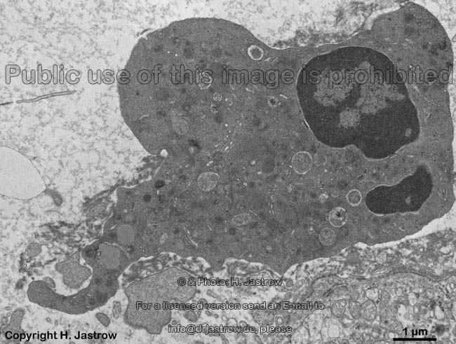
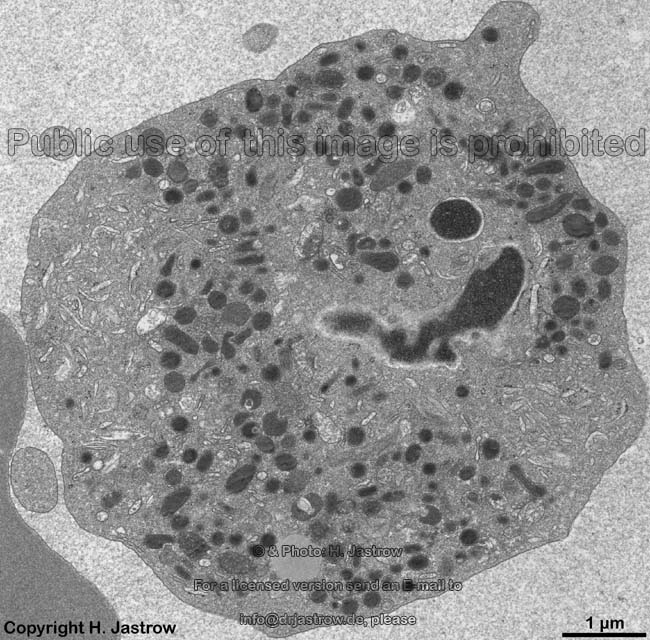
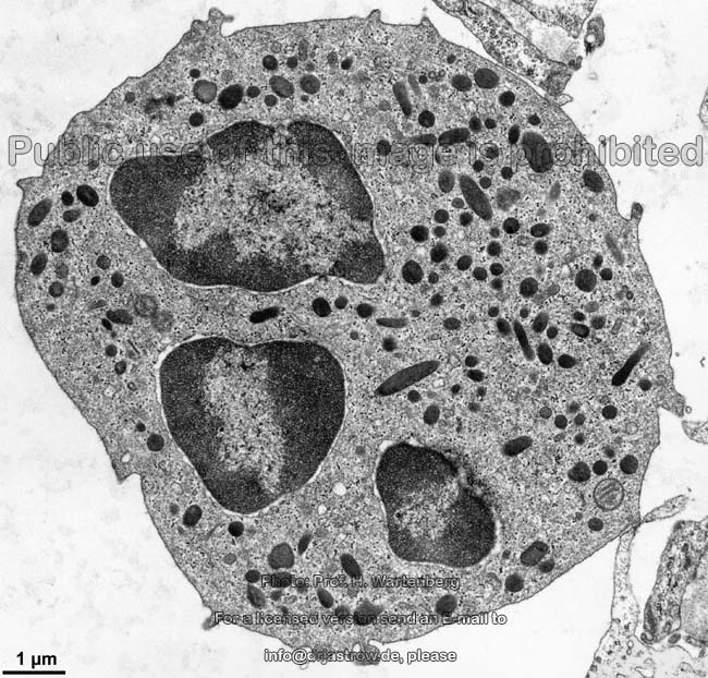
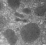
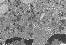
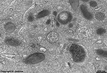
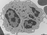
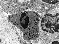
human segmented
neutrophil granulocyte 2 |
detail 1:
cytoplasm |
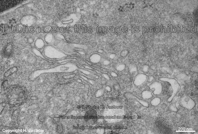
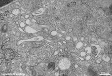
detail 2:
Golgi apparatus |
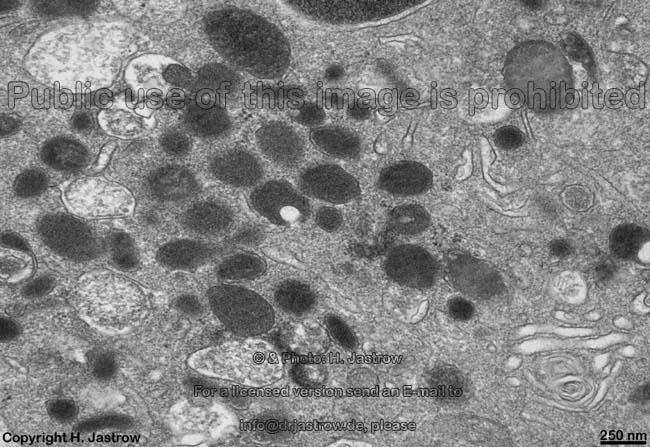
detail 3:
different vesicles |
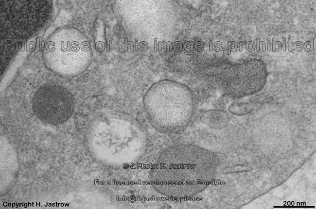
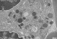
detail 4: specific and
non- specific vesicles |
detail 5:
further vesicles |
|
|
|
|

|
|
detail 6: pore of the
nuclear membrane |
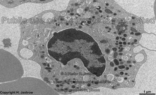
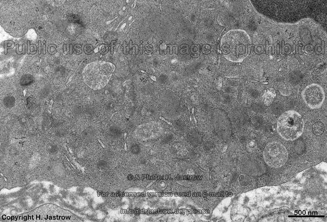
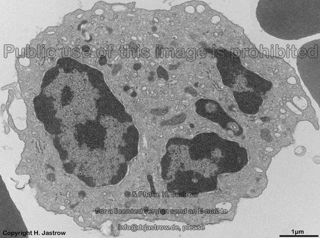
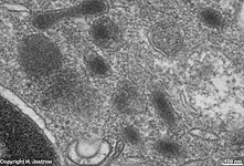
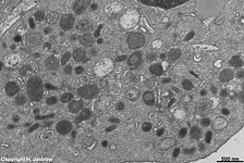
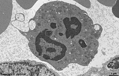
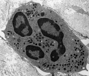
human segmented
neutrophil granulocyte 3 |
detail 1:
cytoplasm + organells |
detail 2: cytoplasm, vesicles,
multivesicular body |
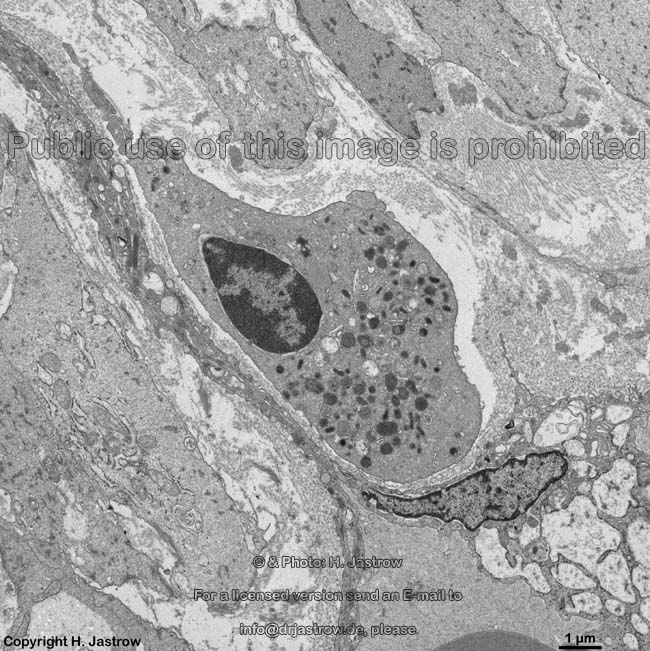
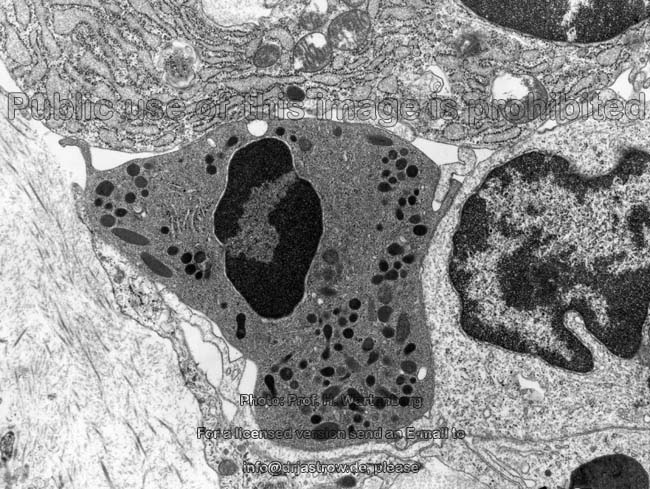
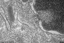
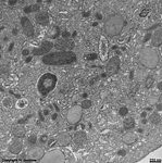
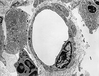
neutrophil in connective
tissue human umbilical cord
|
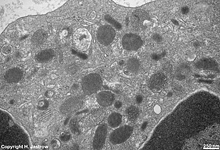
detail: cytoplasm with
organelle |

|
|

|
|
|
|
|
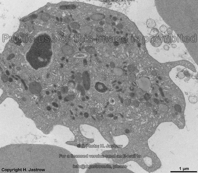
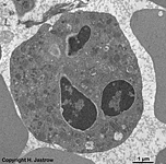
| human segmented
neutrophil granulocyte 4
|
detail: cytolasm
+ organelles |
segmented
neutrophil 5 (human) |
human segmented
neutrophil 6 |
human segmented
neutrophilic granulocyte 7 |
human segmented neutrophil
8 (formalin fixation) |
human segmented
neutrophil granulocyte 9 |
|
|
|
|
 |
segmented neutrophil
granulocyte (monkey) |
segmented neutrophil, plas-
ma cell, lymphocyte (monkey) |
segmented neutrophil
granulocyte (monkey) |
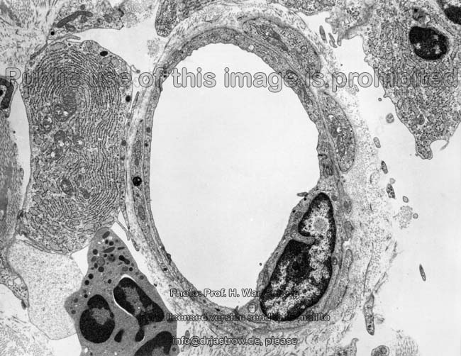
neutrophil close to
a venole (monkey) |
segmented neutrophil
vagina (pig) |
Neutrophilic granulocytes (neutrophils, segmented neutrophilic
granulocytes; Terminologia histologica: Granulocyti neutrophili, Neutrophili,
Granulocyti neutrophili segmentonucleares) belong to the white blood cells
(leucocytes; Terminologia histologica:
Leucocyti) of which they comprise the majority, i.e. 50 to 75 %
in differential blood count. Neutrophils are microphages and thus are capable
to incorporate foreign materials via phagocytosis.
Morphology:
Neutrophils are about spherical when circulating in blood,
in the connective tissue, however,
they appear somewhat longer and ovoid. When cut at maximal diameter
they have a width of 9 to 12 maximal 15 µm.
Under stimulation plenty of elongated pseudopods
form from the cell membrane extending
some hundred nanometres. The nuclei of neutrophils
are rich in heterochromatin resulting
in an intense basophily making it hard to distinguish nucleoli
in light microscopy. Due to the considerable variations of nuclear morphology
the term polymorph nucleus has been established for neutrophils.
Like the other blood cells neutrophils derive
from stem cells and progenitor
cells of the bone marrow from where they
migrate into the blood. After this release
the still
young neutrophils show bend rod-like nuclei
thus the juvenile neutrophils (neutrophilic metamyelocytes; Terminologia
histologica: Metamyelocyti neutrophili, Granulocyti neutrophili juveniles)
are also called
staff cells, Schilling's band cells or band form granulocytes
(Pappenheim stained histological image). They
usually comprise about 5 to 10% of all neutrophils. After some hours the
nuclei begin segmentation, i.e. show larger and smaller lumpy parts interconnected
by very thin bridges. Thus the cells are mature transform into
segmented neutrophilic granulocytes (Pappenheim stained histological
image). This mature form of granulocytes constitutes about 45-74%
of all leucocytes in differential
blood count of adult humans. In case of
purulent
infections this value increases to often above 80% of all leucocytes. If
the relation of staff cells to segmented neutrophilic granulocytes changes
this is called leftward shift in case of >10% staff cells
(typical for acute infections). In
case of < 5% of staff cells we talk about a rightward shift (typical
for e.g., vitamin B12 deficiency anaemia).
segmented neutrophilic granulocytes
are cells of diameters of
10 - 15 µm with 3 - 4 nuclear
segments interconnected via ~100 nm thin nuclear bridges. In
case of too low blood serum levels of vitamin B12 of folic acid a so called
hypersegmentation
of the nuclei gets evident, i.e. nuclei show 5 or more segments.
Neutrophils may also be used for sex
determination: If over 6 of 500 neutrophils (>3%) show a sex
chromatin (Terminologia histologica: Chromatinum sexuale) it is sure that
a female was the blood donor. This appendage of the nucleus, commonly
called drumstick, has a diameter of about 1 - 1,5 µm and comprises
an inactivated X-Chromosome
corresponding to Barr's body of other
cells. In the electron microscope the drumstick reveals as a segmented
very electron-dense part of the heterochromatin.
Besides many other proteins, the cell
membrane of neutrophils expresses the cell surface proteins CD32
(40 KD; an Fc receptor of low affinity for aggregated immunoglobulins)
and CD 68 (110 KD; a mucin-like protein
also known as macrosialin).
An English page with much more detailed information and images is only
available in the professional version of
this atlas
--> Blood cells Overview, eosinophilic
granulocytes, basophilic granulocytes
--> Electron microscopic atlas Overview
--> Homepage of the workshop
Some images were kindly provided by Prof. H. Wartenberg;
other images, page & copyright H. Jastrow.