List of abbreviations
Vocabulary
of micros-
copic
anatomy
specialist terms
explained in
English +
German

Every attempt was made to provide correct information and labelling, however any liability for eventual errors or incompleteness is rejected!

dieser Seite

Editor:
Dr. med.
H. Jastrow


Conditions
of use
Overview stereocilia (Stereocilia):
Pages with explanations are linked to the text below the images when available!
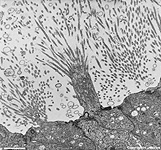
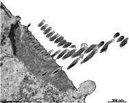
stereovilli of the epididymal
duct (monkey)
stereovilli in the
deferent duct (rat)
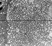
Stereovilli X-section
epididymal duct (monkey)
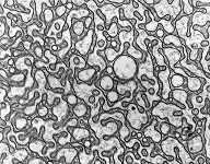
stereovilli X-section 2
epididymal duct (monkey)
outer hair cell stereocilia
organ of Corti (guinea pig)
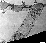
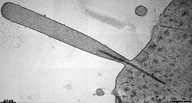
stereocilium = sensory cilia of an inner
hair cell of the ear (guinea-pig)
sensory cilia of an
outer hair cell (guinea-pig)
Stereocilia or stereovilli (Terminologia histologica: Stereocilia; Stereovilli) are immotile long protrusions of the cell membrane.
They contain cytoplasm in which bundles of actin filaments are embedded. The latter are anchored basally to the terminal web and apically to the thickened top of the cell membrane.
There are two different kinds of stereocilia:
1.) sensory cilia of sensory epithelia
Here the long, finger-like stereocilia are seen in rows paralell to each other. They are anchored in a cuticula which is rich in filaments. Stereocilia of inner and outer hair cells of the organ of Corti in the inner ear are about 5 µm in length. Sensory cells of the macula sacculi or utriculi in the labyrith possess cilia which are 5 - 15 µm in length. Type I (bottle-like) and type II (cylindrical) hair cells on the cristae ampullares of the semicircular canals show even longer stereocilia: 5 - 25 µm. In cross sections the round diameter of stereocilia varies from 100 nm at the base to 250 nm slightly above. Sensory stereocilia are similar in construction and appearance to microvilli with the following exceptions: they are longer and greater in diameter, their basis is thinner than the parts located above; a glycocalyx and antennulae are not encountered. Stereocilia belong to secondary sensory cells and change the frequency of generated action potentials depending on direction when being sheared or bend.
2.) fused long microvilli of the epididymal duct or the first third of the deferent duct.
Here stereocilia appear as 3 - 6 µm long fused protrusions of the cytoplasm that have no round but bizarre diameters when cross sectioned. In three dimensions these cilia resemble bizarre coral structures.
In contrast to the sensory stereocilia this kind serves for significant enhancement of the cell surface for better resorption of material from the seminal fluid. The vesicles of the distinct Golgi-apparatuses of their epithelial cells are secreted in the lumen in between the cilia. There are no antennulae or glycocalyx seen on top of these cilia. Actin filaments are not as clearly visible as in microvilli or the other kind of stereocilia.

--> further cell surface differentiations: kinocilia, microvilli, sensory epithelia
--> Electron microscopic atlas Overview
--> Homepage of the workshop


Some pictures were kindly provided by Prof. H. Wartenberg; other images, page & copyright H. Jastrow.