Overview colon (Colon):
Pages with explanations are linked to the
text below the images if available! (Labelling is in German)
 |
 |
 |
 |
 |
 |
 |
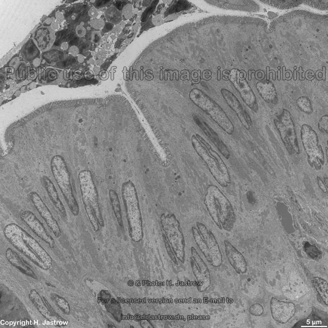
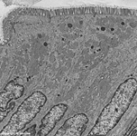
lumen, epithelial cells
colon (rat) |
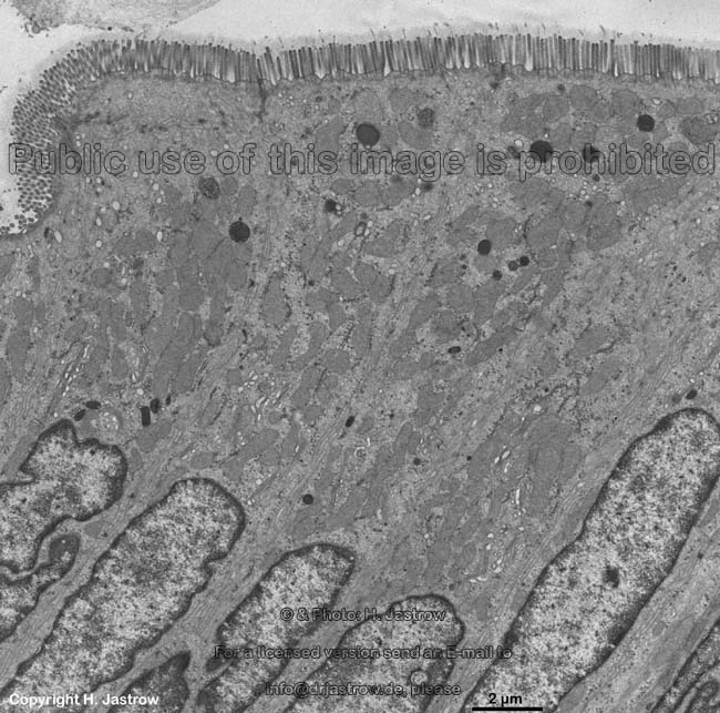
apical cytoplasm
resorbing cells (rat) |
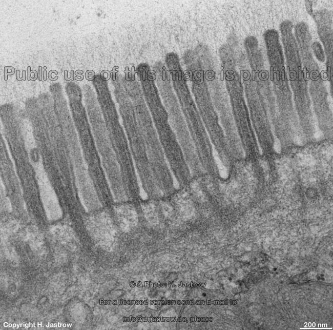
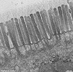
microvilli + glykocalix
enterocyte (rat) |
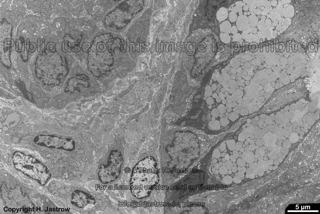
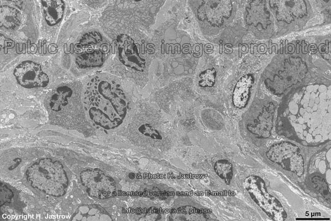
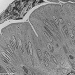
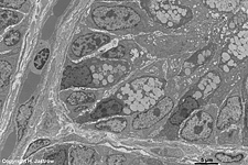
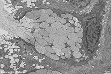
base of a crypt with
many goblet cells (rat) |
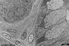
goblet cells, Lamina propria
(rat) |
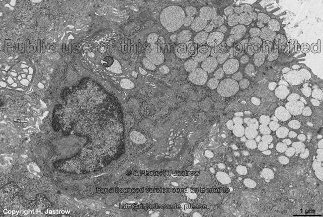
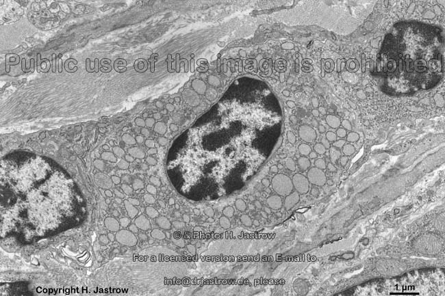
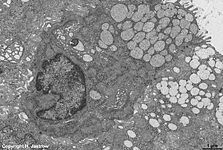
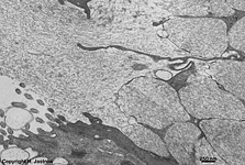
goblet cell (rat) |
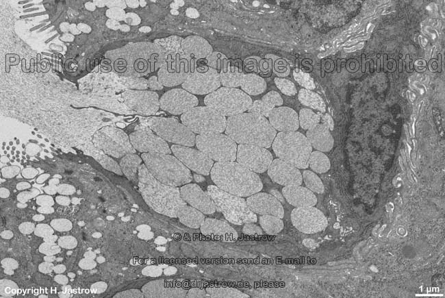
secretion of a goblet cell
(rat) |
 |
 |
 |
 |
 |
 |
detail thereof: secretion
(rat) |
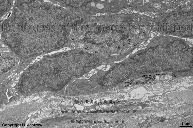
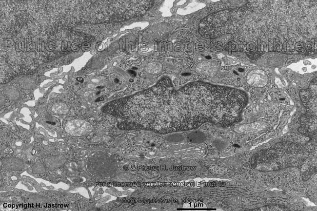
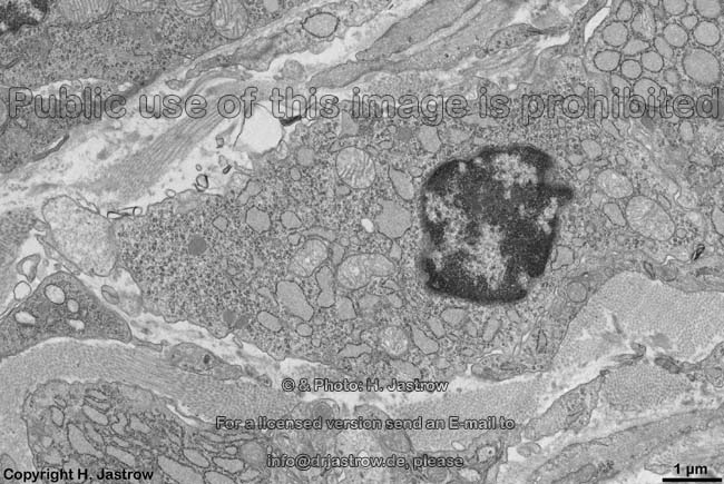
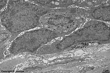
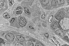
base of a crypt with 2
enteroendocrine cells (rat) |
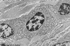
detail: enteroendocrine cell
(rat) |
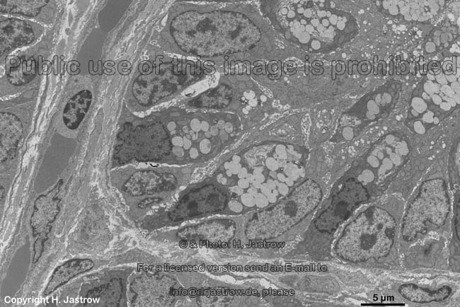
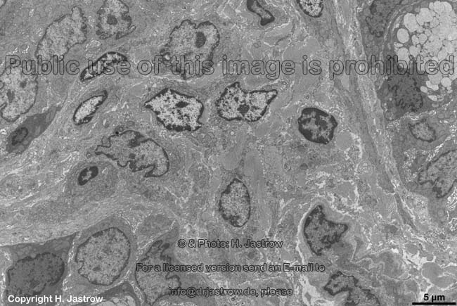
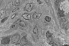
Lamina propria mucosae with
connective tissue cells (rat) |
idem (rat) 2 |
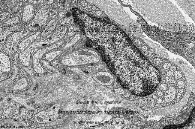
plasma cells in the
Lamina propria mucosae (rat) |
The colon (Terminologia histologica: Colon) is the major
part of the large intestine
and has a length of 1,3 to 1,5 m and a diameter which is about 3 times
that of the small intestine
(duodenum,
jejunun
+ ileum). The colon has several segments and
begins after the caecum (Terminologia
histologica:
Caecum) in form of
the retroperitoneal ascending colon (Colon ascendens). After the
right
flexure of the colon (Flexura coli dextra) it is followed by
the intraperitoneal transverse colon (Colon transversum) which after
the left colonic flexure (Flexura coli sinistra) continues
as the retroperitoneal descending colon (Colon
descendens) to become intraperitoneal in the pelvis as sigmoid (Colon
sigmoideum). The rectum finally is the
last part of the large intestine.
The colon has 3 longitudinal stripes (taeniae
coli;Terminologia histologica: Taeniae coli termed: Taenia libera,
- mesocolica & -omentalis) in which the longitudinal layer of the muscularis
forms strong bands (see below). Fatty appendages
(Appendices epiploicae) are attached
to the free taenia (Taenia libera). They mainly consist of loose connective
tissue and unilocular fat cells.Haustrae
are protrusions of the colon located between incisions visible from outside
that from inside appear as semilunar
folds of the mucosa (Plicae semilunares). These plicae semilunares
are no permanent folds but they disappear when the local contraction of
the circular muscle layer which is
the cause of their existence diminishes.
The colon (Terminologia histologica: Colon) is characterised
by
exclusively crypts and a large diameter. Its wall is similar
to gut in general but has some specialities. The mucosa
has a superficial
Lamina epithelialis bordering the lumen with vast
amounts of goblet cells,
enterocytes
and a few enteroendocrine cells at the base of
the crypts. The underlying loose connective tissue
layer is called Lamina propria mucosae. It shows very large aggregations
of secondary lymph follicles that join each other
(Folliculi lymphatici aggregati = Payer's plaques), that may reach
down in further layers. A thin layer of smooth
muscle cells (Lamina muscularis mucosae) is the deepest layer
of the mucosa. The following
Tela submucosa is a thicker layer of loose connective
tissue which shows the submucous nerve plexus
(Plexus submucosus; Meissner's plexus) and extends to the following
Tunica
muscularis. The latter has a circular layer (Stratum circulare)
which is very thin in the area of haustres and a logitudinal layer (Stratum
longitudinale) which is only evident in the region of taenia. In between
these two smooth muscle cell layers the Plexus myentericus (Auerbach-Plexus,
a further autonomous nerve plexus of the gut)
is encountered. The outermost layer Tunica
serosa is a small loose connective tissue
layer (Tela subserosa) covered with the monolayered squamous epithelium
of the peritoneum only in areas where the colon lies intraperitoneally
(e.g., transverse and sigmoid portion). In other (retroperitoneal) regions
an adventitia formed by connective tissue
anchors the colon to neighbouring structures.
--> Table of enteroendocrine cells
--> ileum, plasma
cells
--> Electron microscopic atlas Overview
--> Homepage of the workshop
Images, page & copyright H. Jastrow.