
cross-sections at highest resolution
Dr. H. Jastrow
Department of Anatomy, Histology, Johannes Gutenberg-University,
Mainz, Germany
This presentation was shown at the 4th Visible Human Conference at Keystone, Colorado U.S.A. on October 19 2002.
|
cross-sections at highest resolution Dr. H. Jastrow
This presentation was shown at the 4th Visible Human Conference at Keystone, Colorado U.S.A. on October 19 2002. |
|
Fig.1
 |
Ladies and gentlemen,
The rescanned visible human male sections have a resolution which is much better than that of the first data set. They serve as basis for the atlas which I am going to present now. Fig. 2 shows the material used: the atlas is based on all rescans of the original films taken from the visible human male during sectioning. The data was downloaded from the FTP-server of the NLM. Further all CT and MR-images were obtained from different mirror sites. |
Fig.2
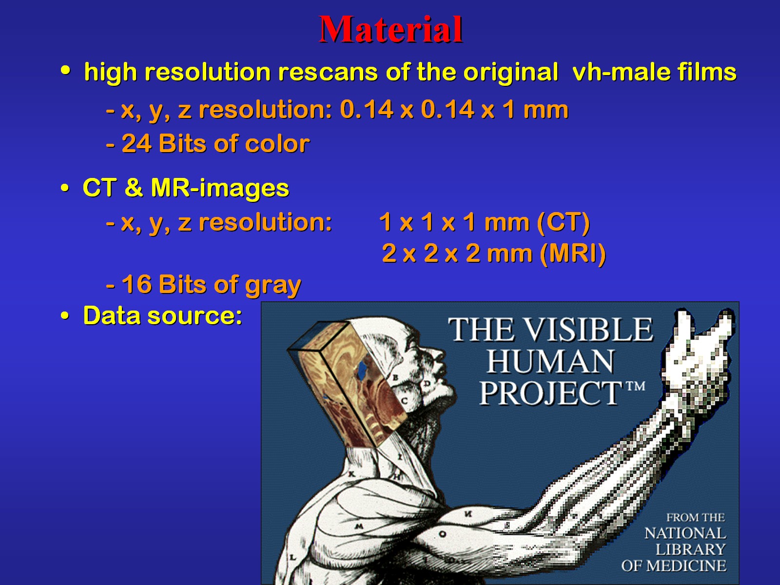 |
Fig.3
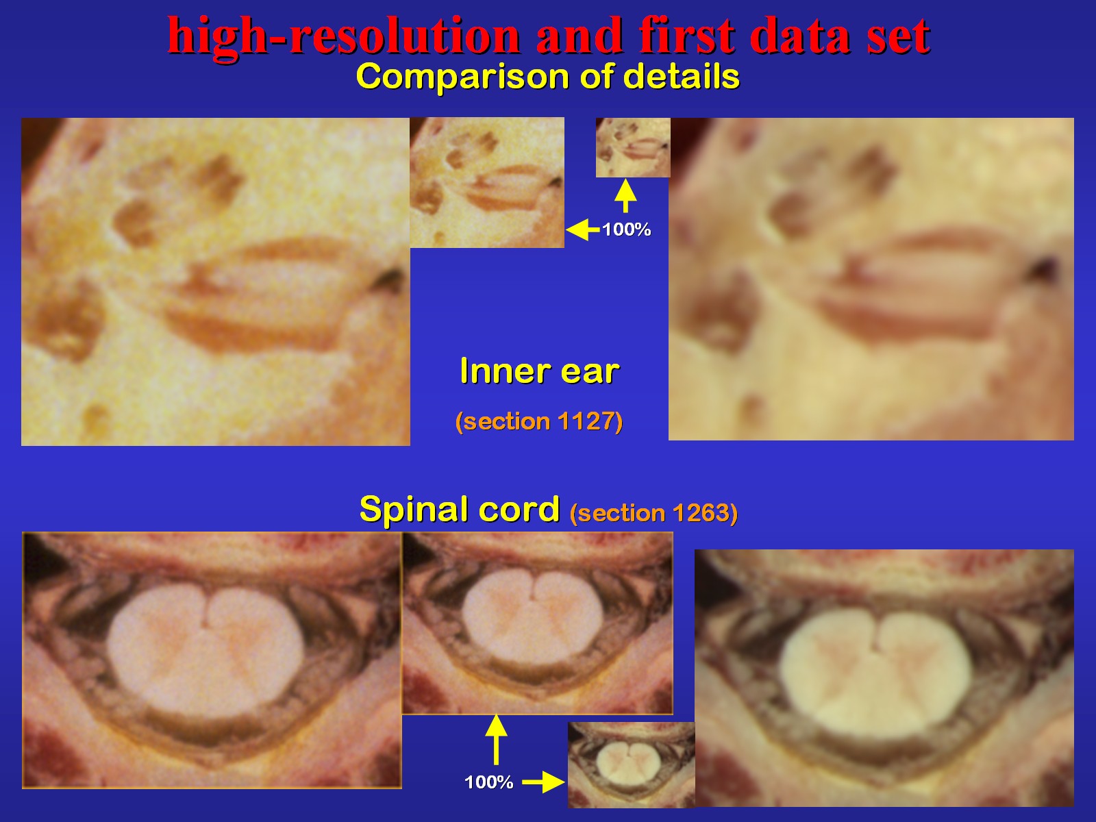 |
The resolution of the rescanned images is nearly
2.2 times higher than that of the first data set. This is evident when
regarding details like the ones shown in Fig.3.
After decompression the rescanned sections appear similar to the one shown in Fig.4. It is now used to demonstrate you the processing of the images used in the present atlas. |
Fig.4
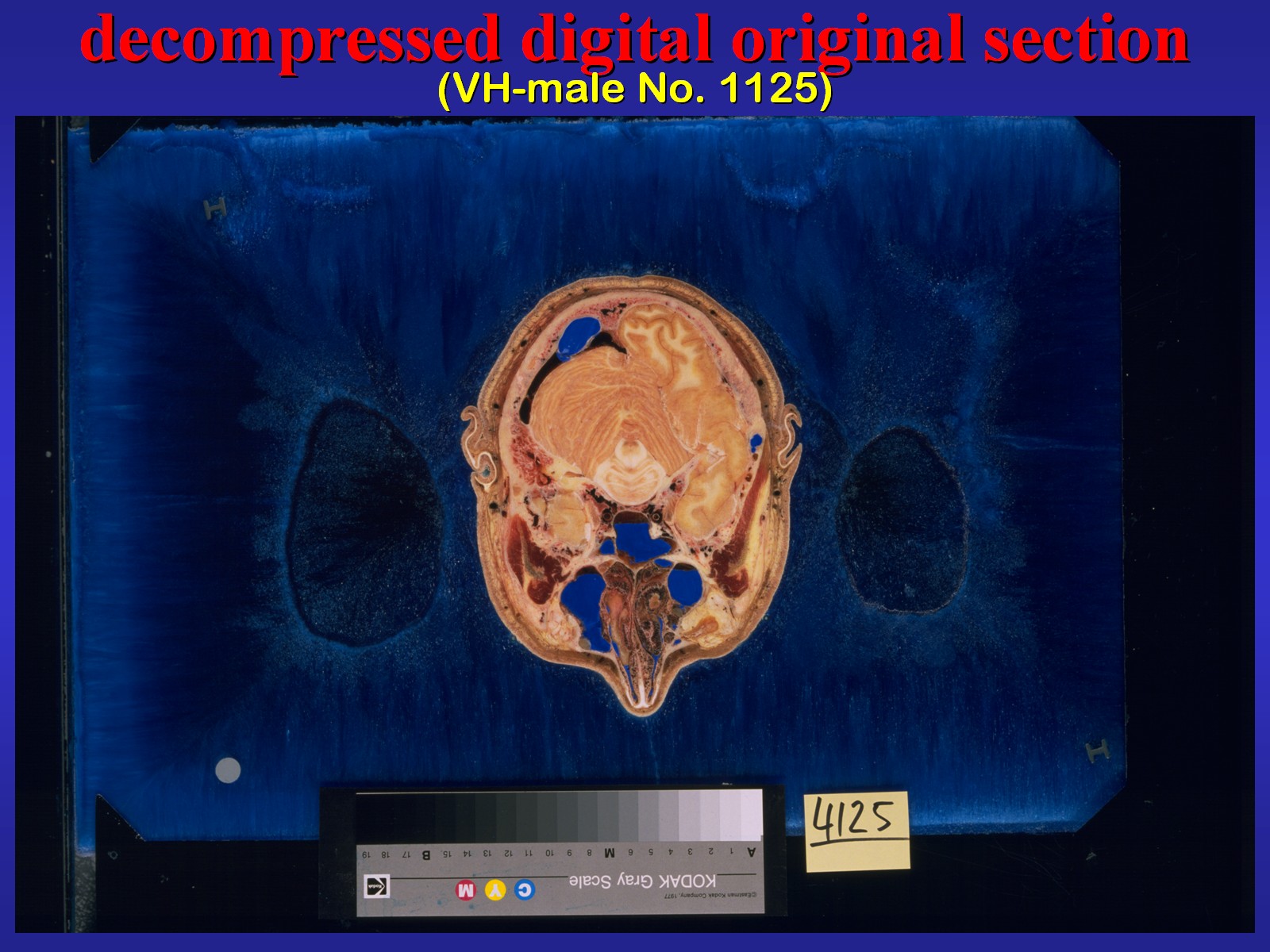 |
Fig.5
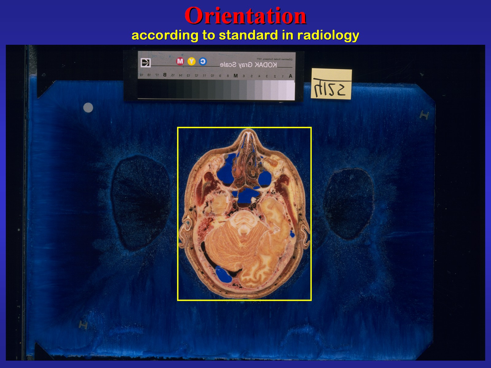 |
After orientation according to the standard established
in radiology images were trimmed (Fig.5).
Thereby the original resolution was maintained. Then a black background was created and edges were cut sharp (Fig.6). |
Fig.6
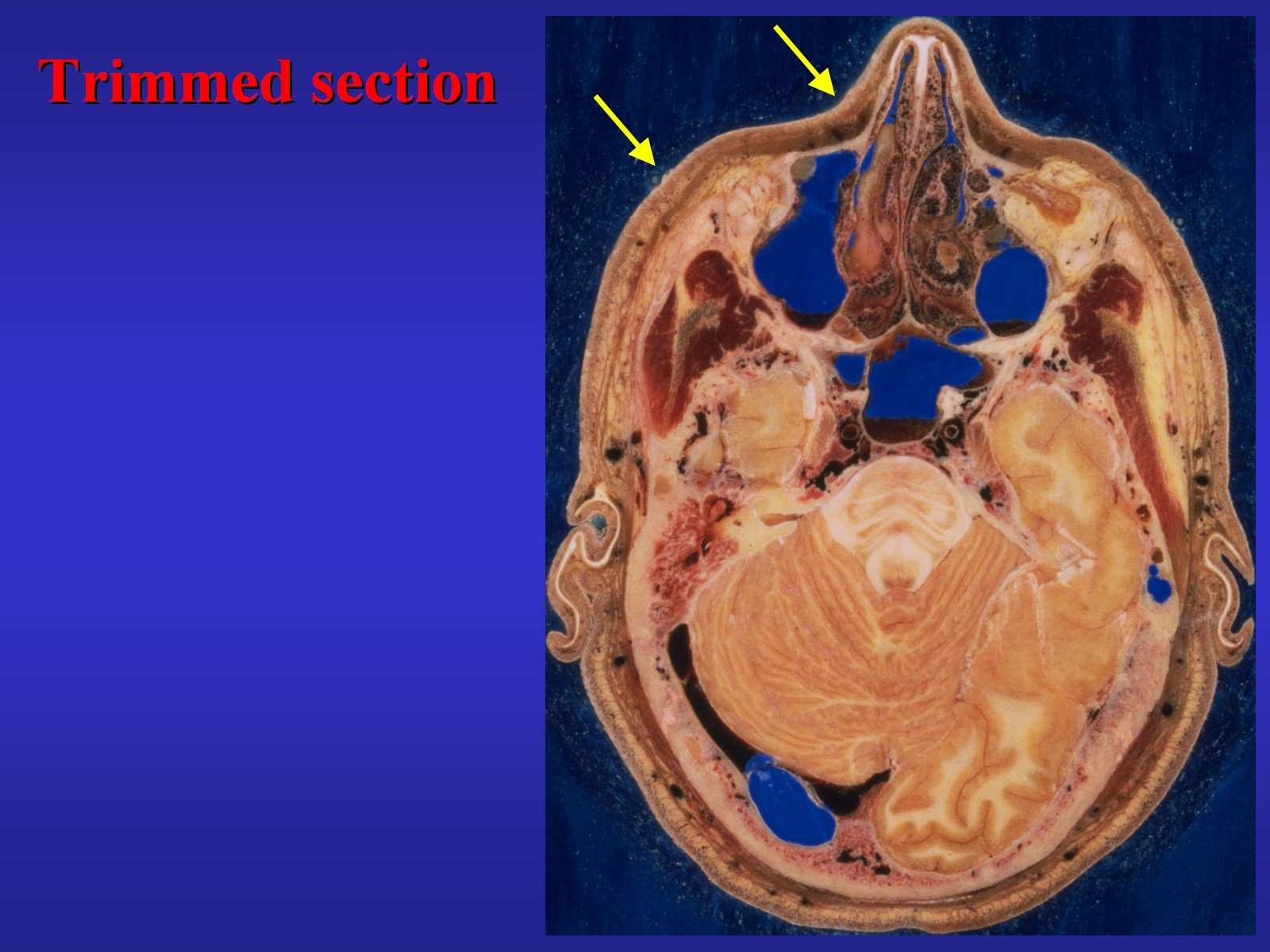 |
Fig.7
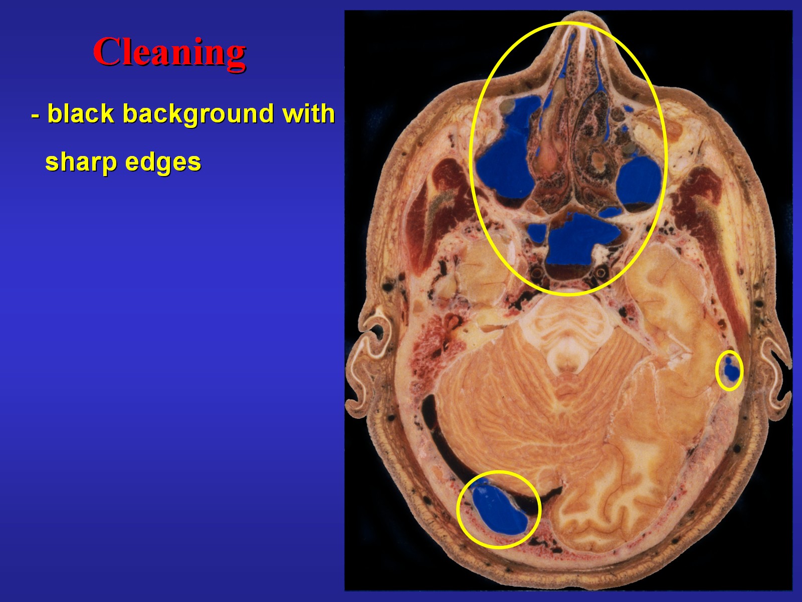 |
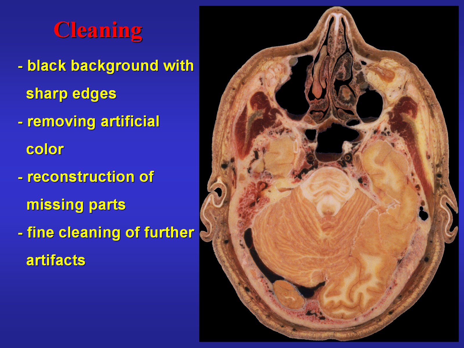
As you can see in Fig.7 the blue artificial color filling of spaces inside the sections was removed (Fig.8). Mucus of the air ways was erased and parts of the sections that were destroyed during the cutting procedure were reconstructed using image information from the neighboring sections. Any further artifacts were removed as far as possible from all images. | Fig.8
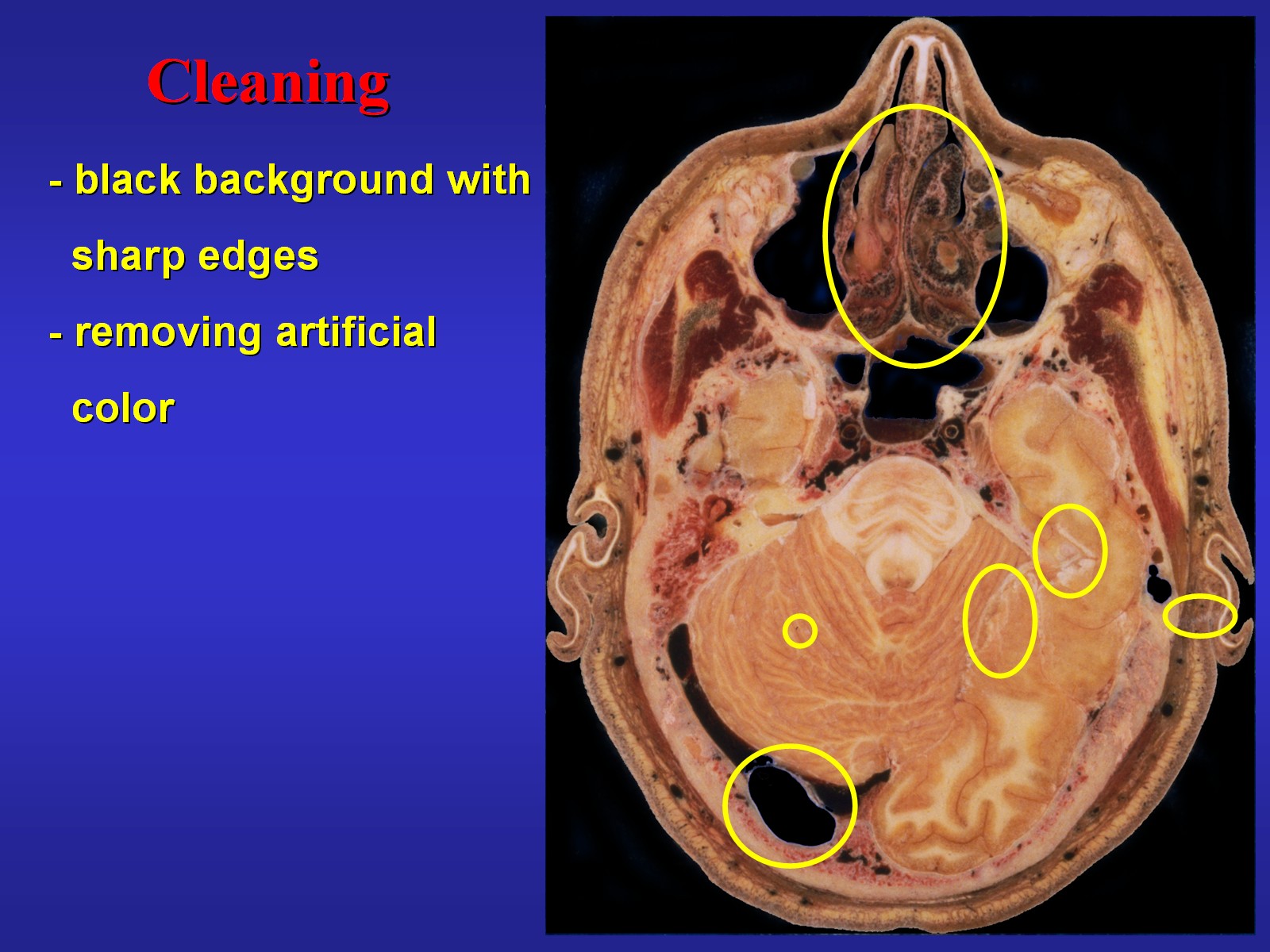 |
Fig.9
 |
This was done with a small brush superimposing pixels from places with a color and texture similar to that of the artifact region (Fig.9). This accurate fine cleaning was the most time-consuming part of the image processing. Finally, a color adjustment with channel-wise optimization was applied to make images more brilliant (Fig.10). | Fig.10
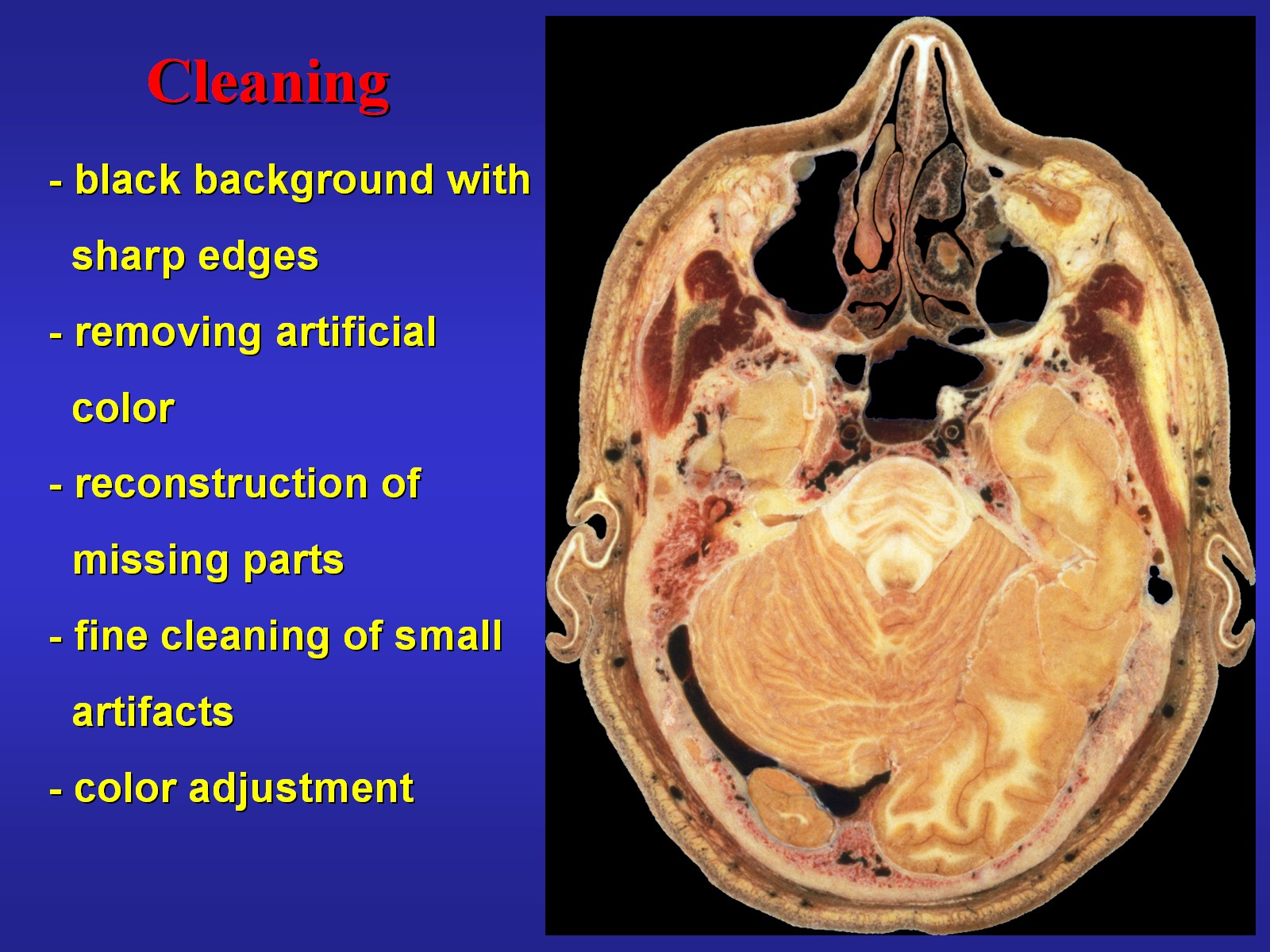 |
Fig.11
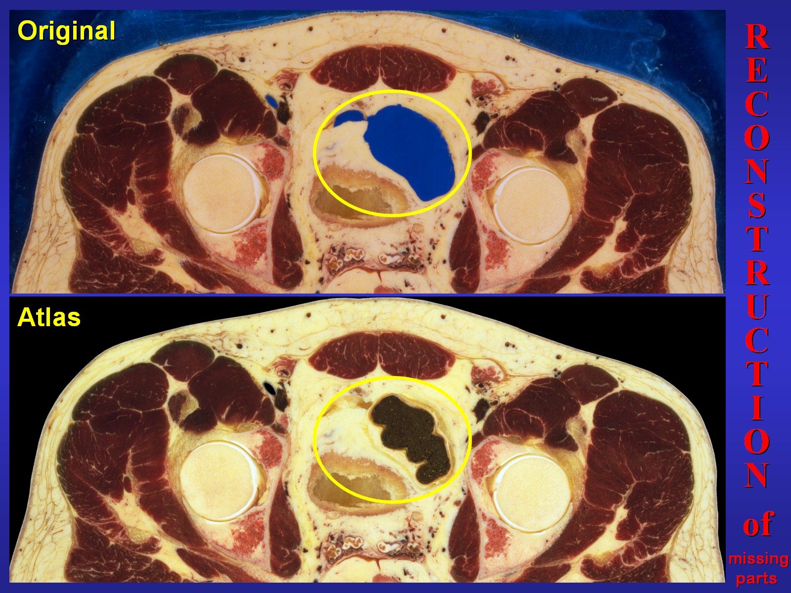 |
On the example of a section of the pelvis (Fig.11) I demonstrate you the reconstruction of missing image information. We are going to look at the marked region now. Fig.12 shows the steps: at first the background was exactly overlaid from the closest sections where it was preserved. Then the missing part of the gut with surrounding fat was copied in along the edges. This was done in many small steps leaving small portions out since the copy source was larger than the area to be filled. Further the outline of the gut was corrected regarding changes between the sections up to the source section which was the last one showing the colon. | Fig.12
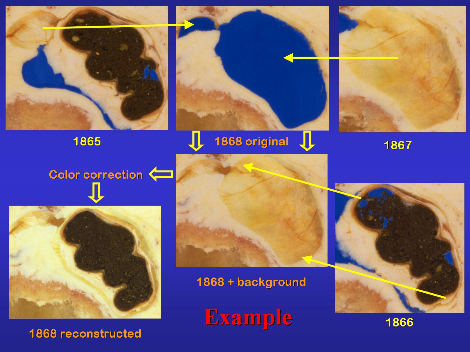 |
Fig.13
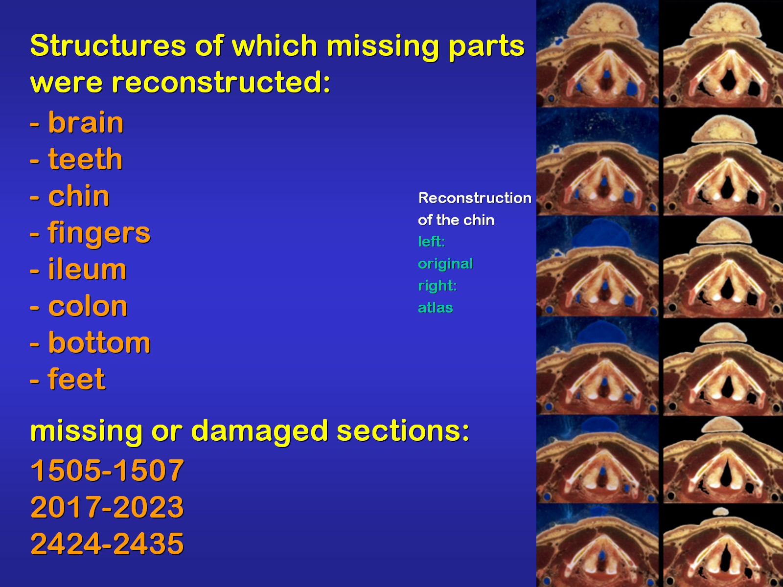 |
The list in Fig.13 shows which missing
parts of the original data were reconstructed to produce sections of the
complete visible human male. Of course one has to be aware that even the
most accurate reconstruction, which was tried here, cannot exactly repair
structures destroyed during sectioning.
CT and MR-images were converted from 16 Bits of gray to 24 Bits of color. Artifacts were removed. For the non-axial MRI, which was the majority, the axial plane was calculated using a software (Fig.14). |
Fig.14
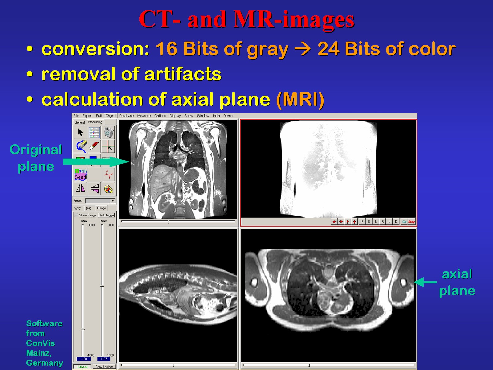 |
Fig.15
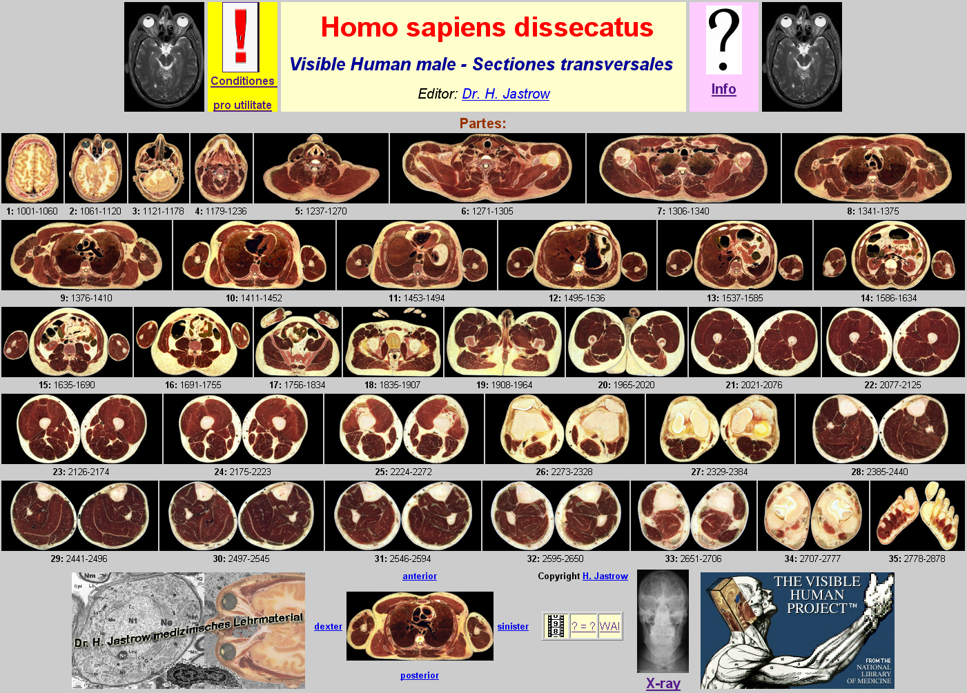 |
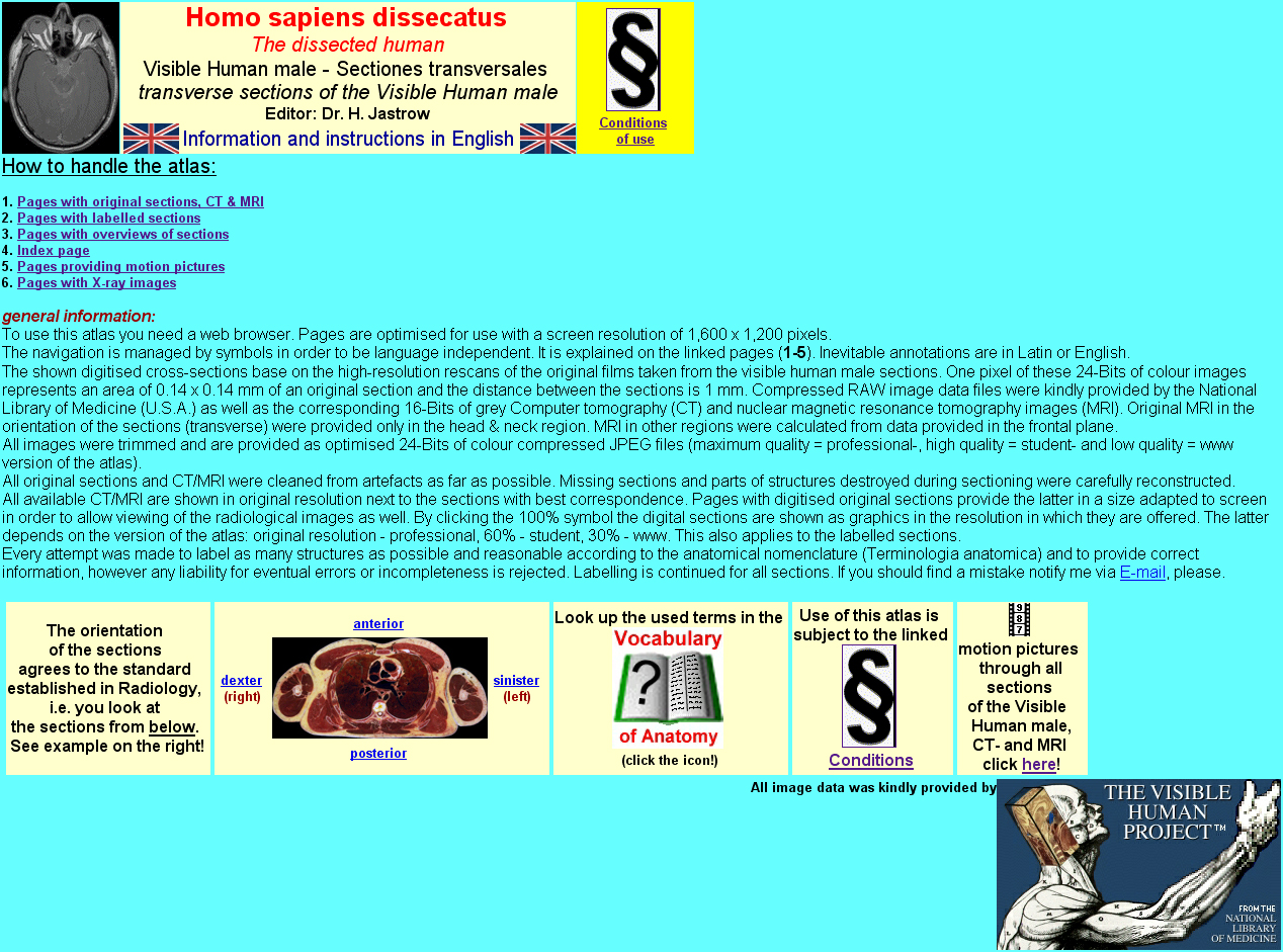
Fig.15 shows the index page of the atlas.
The right upper icons lead to conditions of use and information pages.
The small images each represent the portion of sections whose numbers are
given below. They are linked to section overview pages. Further down image
orientation is shown. The three small icons left to the VHP-Logo lead to
motion pictures, a vocabulary of gross anatomy and the homepage of the
workshop anatomy for the internet providing the web version of this atlas.
Further several X-ray images are available on a page linked to the x-ray
icon.
The info page provides links to instructions in 4 different languages (Fig. 16). |
Fig.16
 |
Fig.17
 |
When choosing English, the page shown in Fig.17
providing general information is loaded. It is linked to pages explaining
navigation and handling of the different kinds of pages of the atlas.
The first of these pages (Fig.18) explains the page layout and the symbols of the navigation bar. Under the example page on the right you see an enlargement of such a bar. It was designed to make navigation easy and language independent. I now demonstrate you the navigation on an example. |
Fig.18
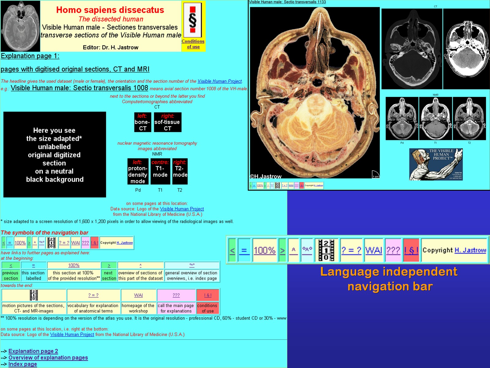 |
Fig.19
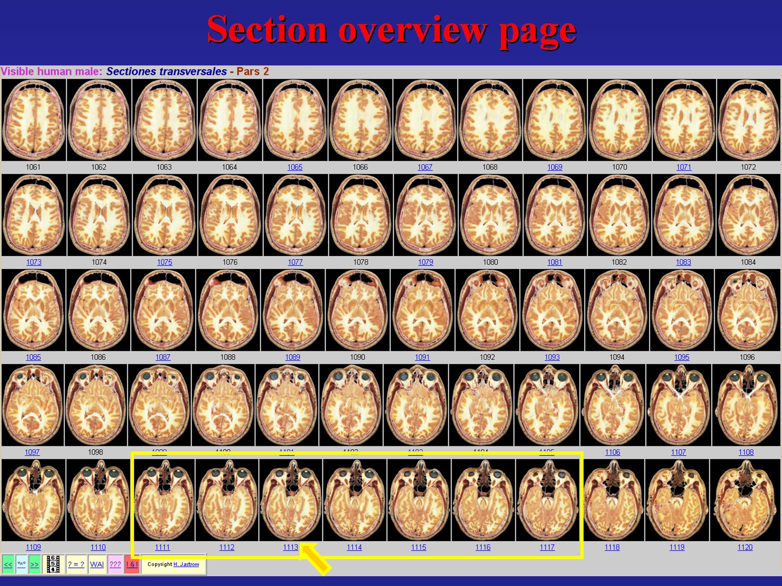 |
Imagine we are looking for a section showing the pituitary. After clicking the second section icon on the already shown index page, we come to this section overview page (Fig.19). The gland can be seen on the marked images. With a click on one of them the page with the digital section, CT and MRI appears (Fig.20). To have a closer look at the structure of interest one only has to click on the hundred percent button. | Fig.20
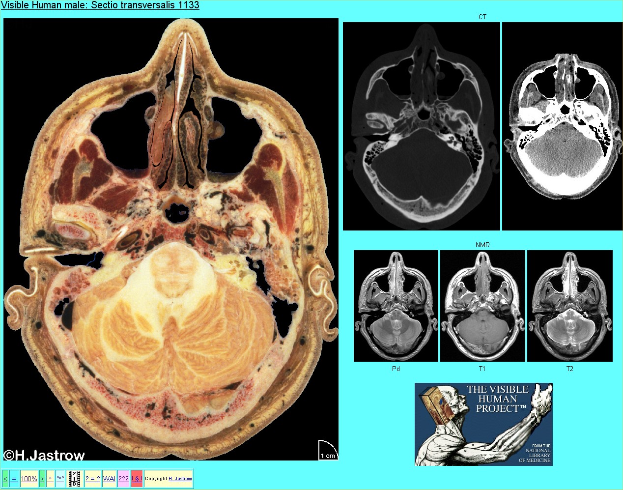 |
Fig.21
 |
To be able to demonstrate the high resolution
of the image called up now just a part of it is shown in Fig.21.
The motion picture of Fig.22 was made from neighboring head sections. It is an example for motion pictures that I can generate from the data set on demand. The ones that can be started from the animation page have not quite the resolution and quality of the one shown at the conference. |
Fig.22
Motion picture reolution dramatically scaled down here. The original shown at the conference had a file size of 768 MB! |
Fig.23
 |
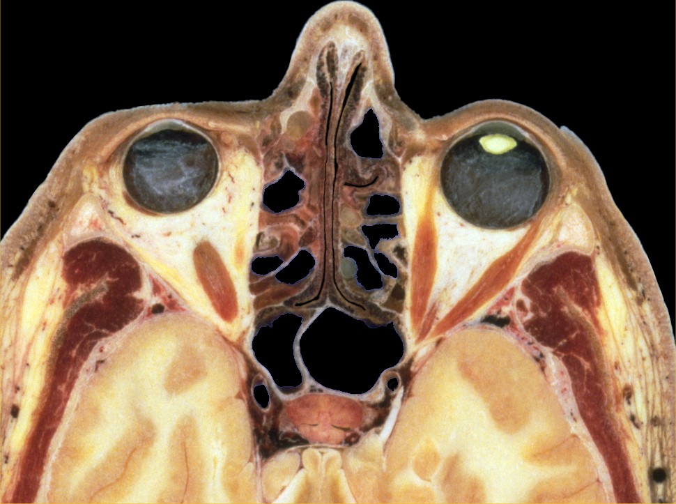
At present, more than 200 sections are labeled
in great detail. Some of these images did not provide enough space to give
all terms of interesting regions. In such cases (Fig.23) labeling
is provided on neighboring sections or on a separate detail page.
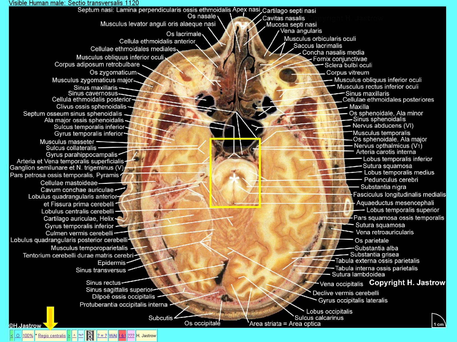
Fig.24 is an example of such a labeled detail page. It shows the central region of the previous image with the brain-stem. |
Fig.24
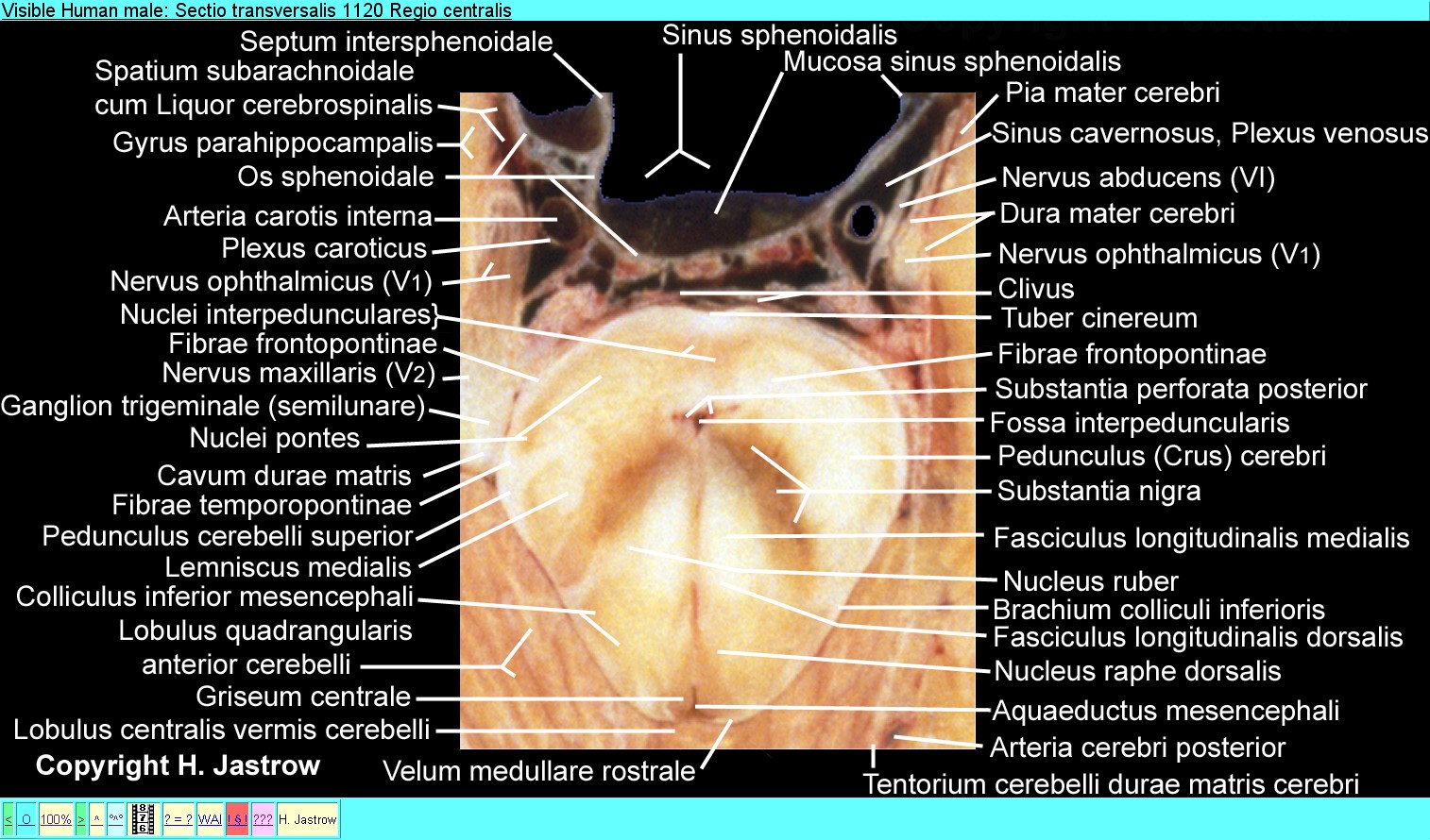 |
Fig.25
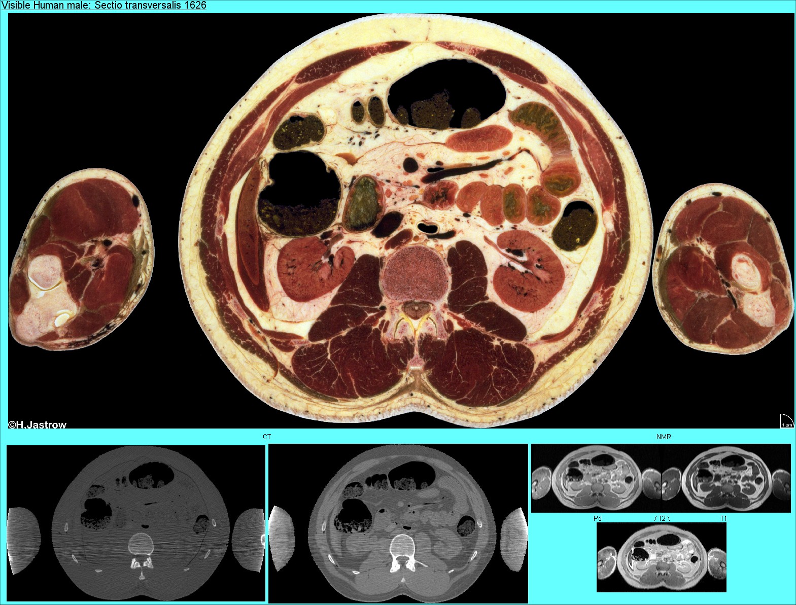 |
Fig.25 demonstrates a page of the abdomen
with calculated MR-images.
Fig.26 is a page of the thorax which also provides reconstructed MRI. |
Fig.26
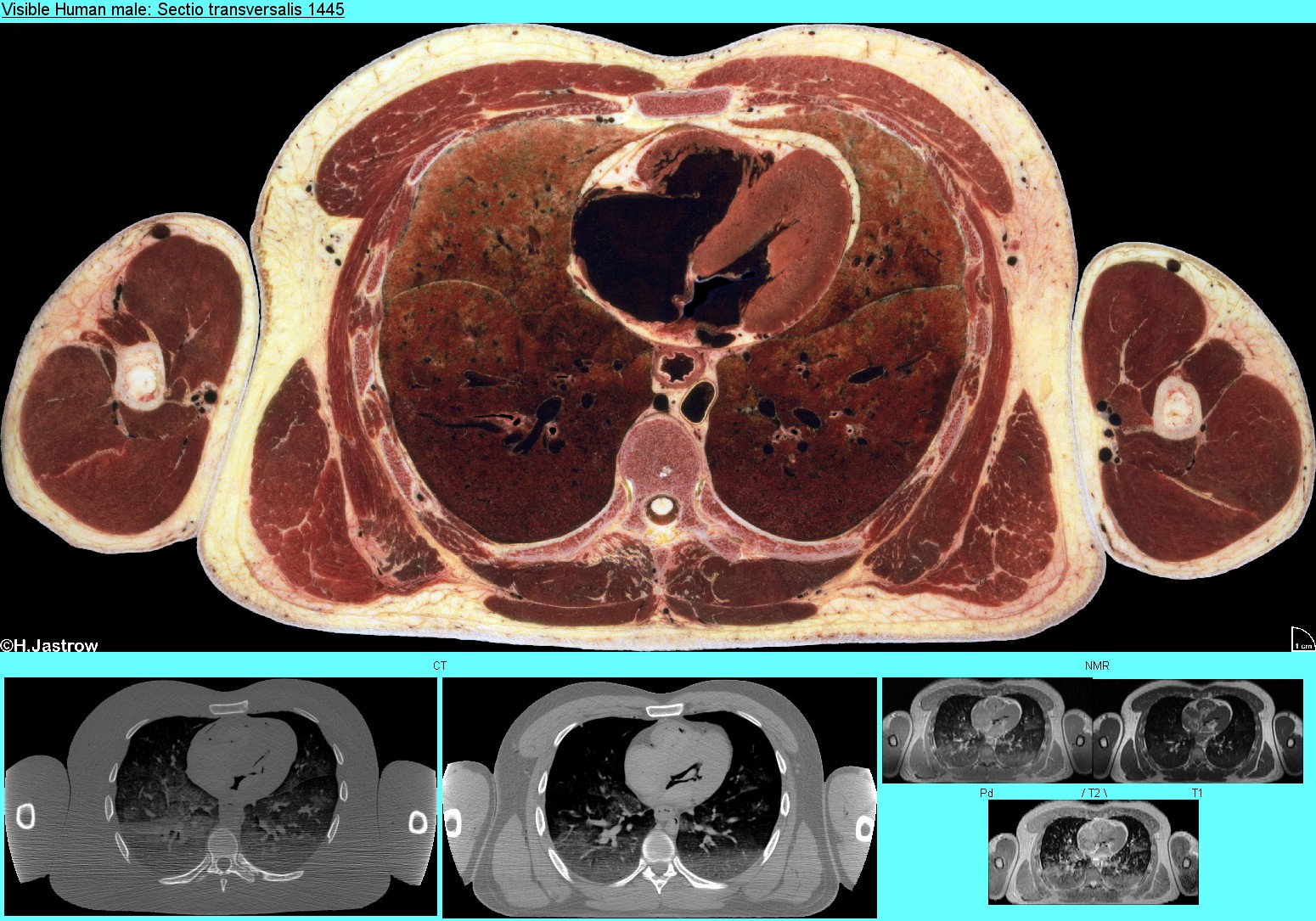 |
Fig.27
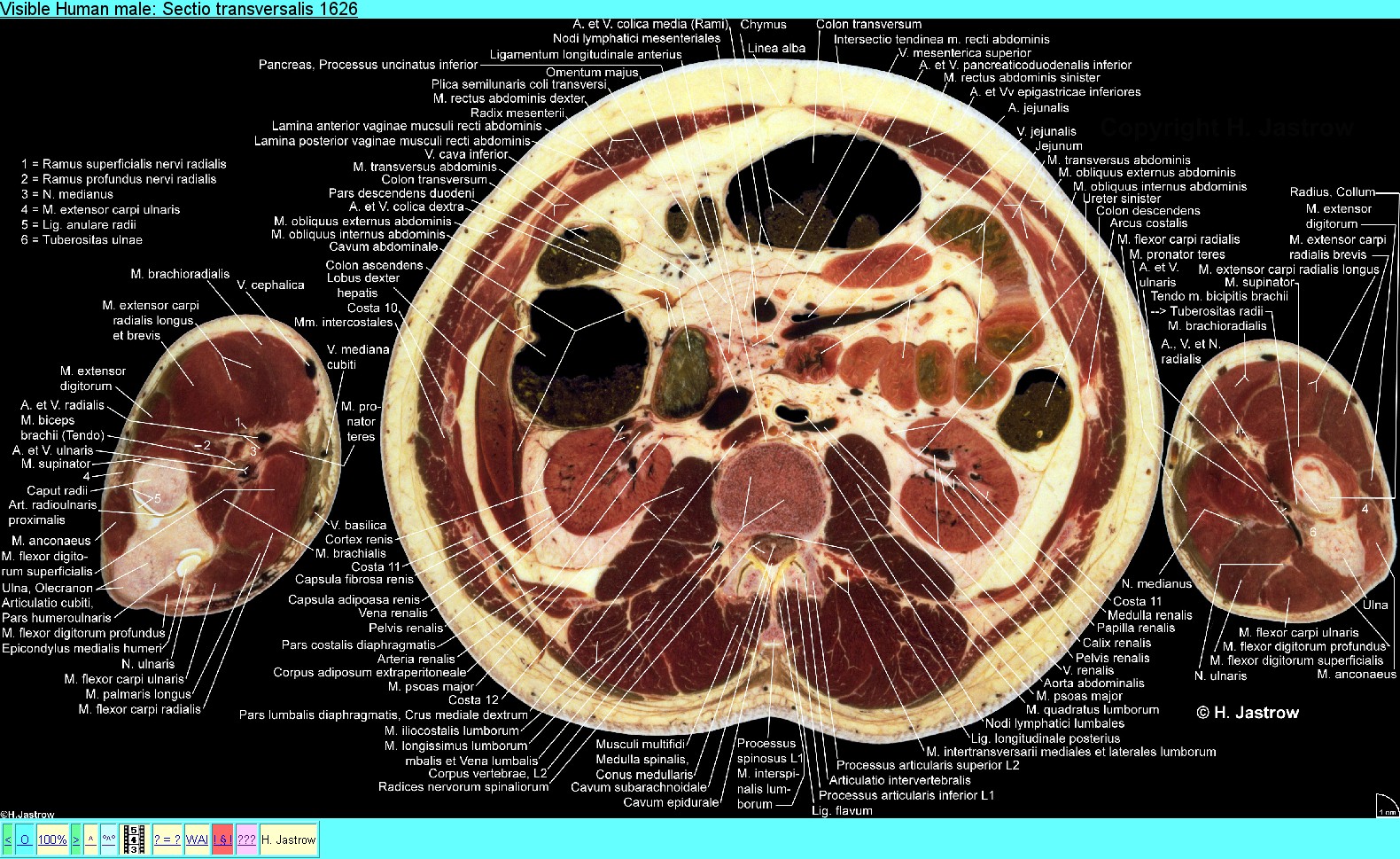 |
Note that there are over 120 anatomical terms in Fig. 27. It
is the labeled version of Fig.25. To maintain clearness, there is
no crossing of the lines.
On the main motion picture page (Fig.28) the different kinds of animations are listed. A detail of one of the six linked overview pages is shown below. At present a total of over a 120 different animations is available. |
Fig.28
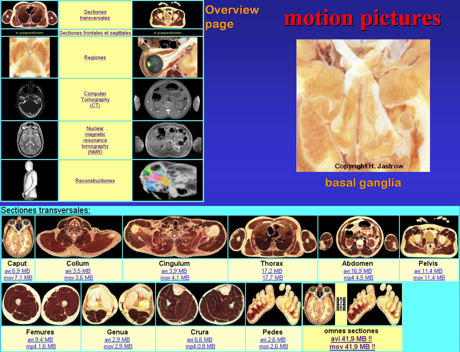 |
Fig.29
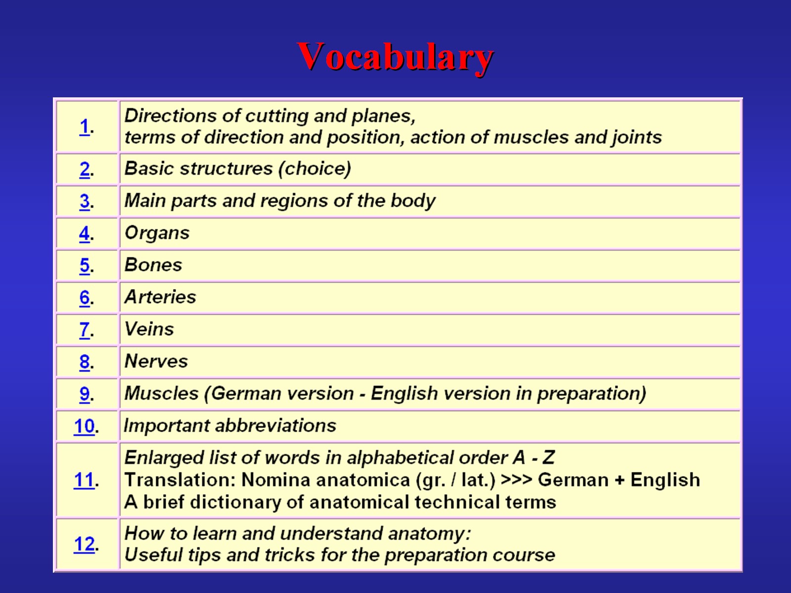 |
The linked vocabulary of gross anatomy (Fig.29) provides over
850 Latin anatomical terms and their corresponding expressions in English
and German. The terms are arranged according to subject as well as alphabetically.
Thus unclear labelling can be looked up easily.
In Fig.30 you see in how far the three versions of the atlas differ and their planned date for release. All versions include the complete transverse sections and images labelled up to the date of purchase. |
Fig.30
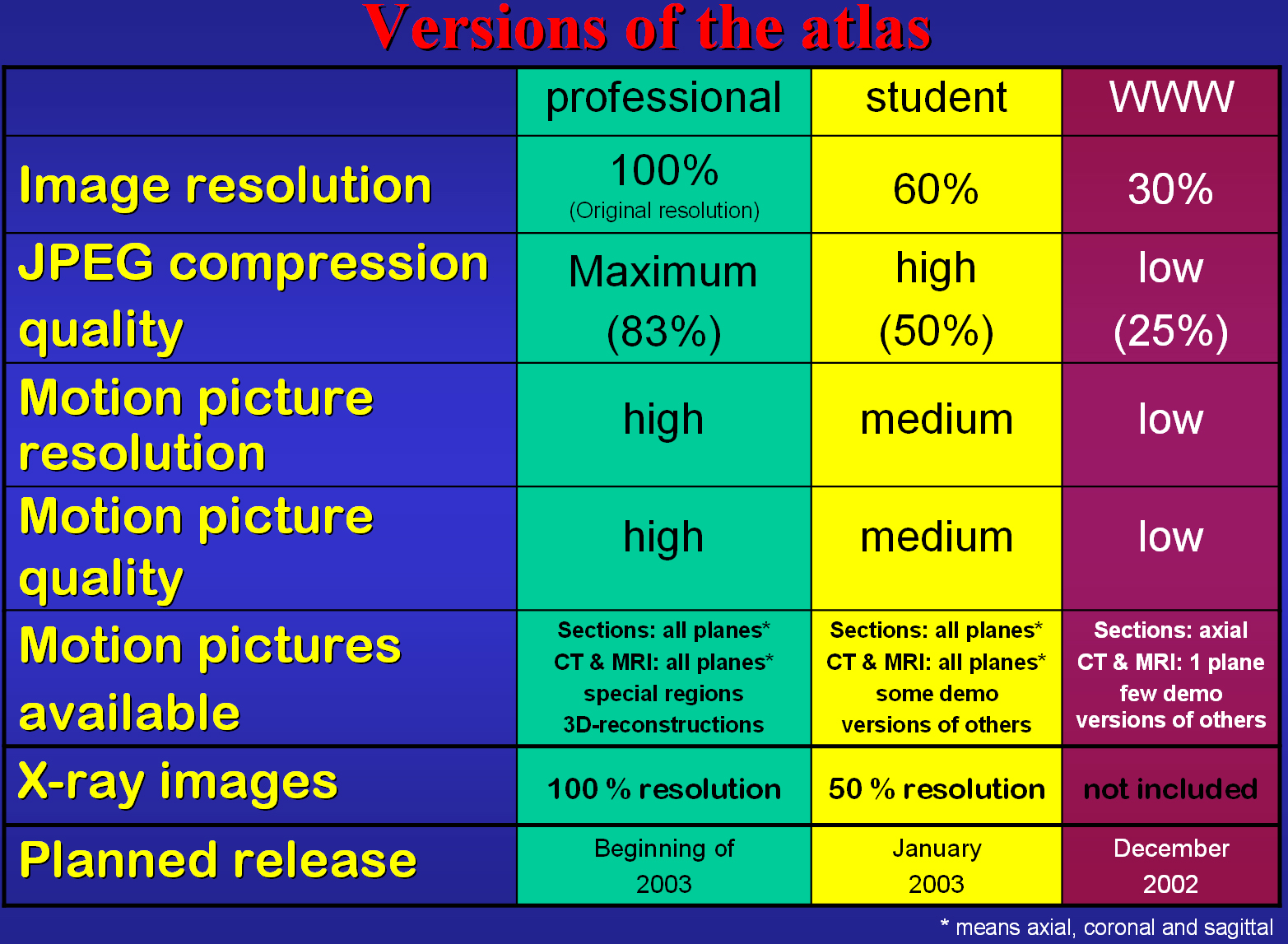 |
Fig.31
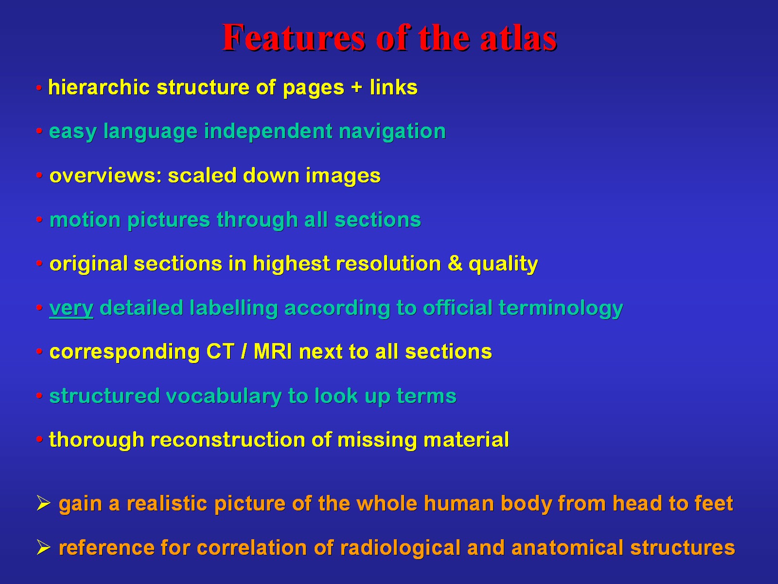 |
By the points listed in Fig.31 the atlas is intended to be a
useful reference for medical education and research. It enables a visual
journey through the whole body showing the complete male human gross anatomy
at the highest available resolution. It contributes to a three-dimensional
understanding of topographic relationships. All important structures of
gross anatomy are thoroughly labelled using the standard terminology to
make the atlas ideal for use everywhere. Anatomy can be directly compared
to corresponding radiological images.
Many people can benefit from the atlas since the material offered can be used for teaching as well as learning gross anatomy in general. The spectrum ranges from university, health care related schools and colleges to lay people interested in the construction of the human body (Fig.32). |
Fig.32
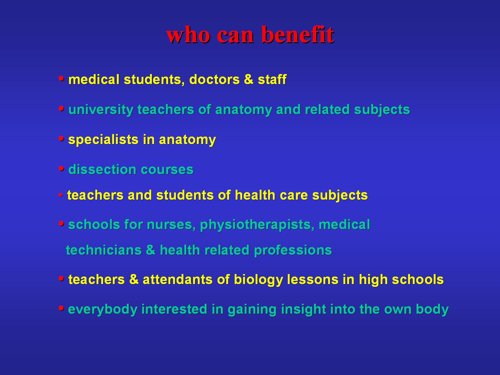 |
Fig.33
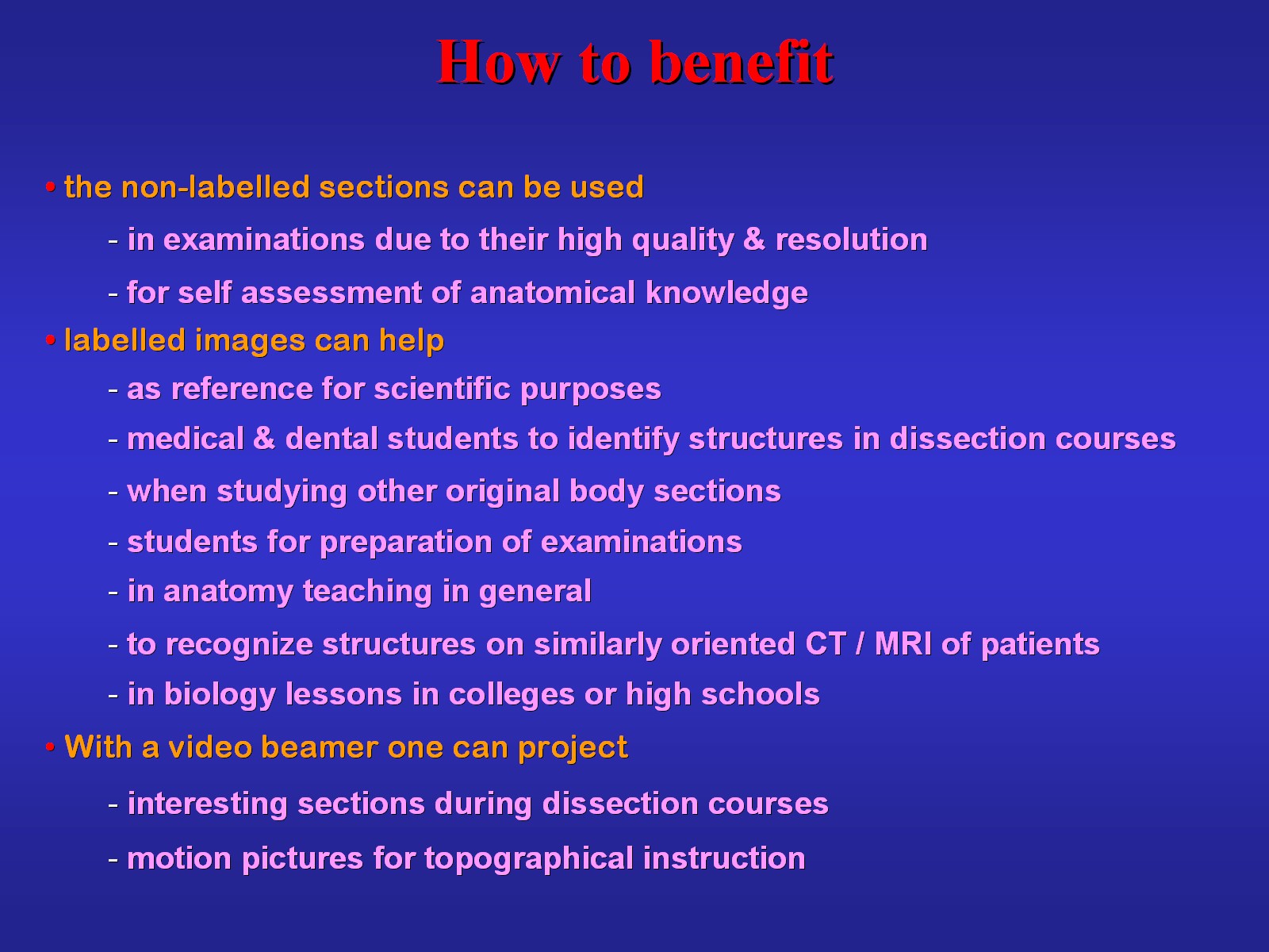 |
Figs.33 and 34 list some examples of how the different
material contained in the atlas can be used for study and instruction of
anatomy. Due to its high quality is suitable as reference for scientific
purposes and appropriate for instruction of specialists and students. It
is a valuable source for medical doctors and staff and also beneficial
for teachings of clinical anatomy as well as biology lessons in college
and high school.
For non-professionals the vocabulary is a valuable key to understanding the international terminology. The motion pictures facilitate acquisition of a 3D-understanding of anatomy and topographical relationships. In the training of medical students and doctors it is essential to correlate anatomical structures of the human body with radiological images. For this reason sections are presented next to corresponding radiological images that also can serve radiologists as a reference. In brief the atlas is a source for many uses. |
Fig.34
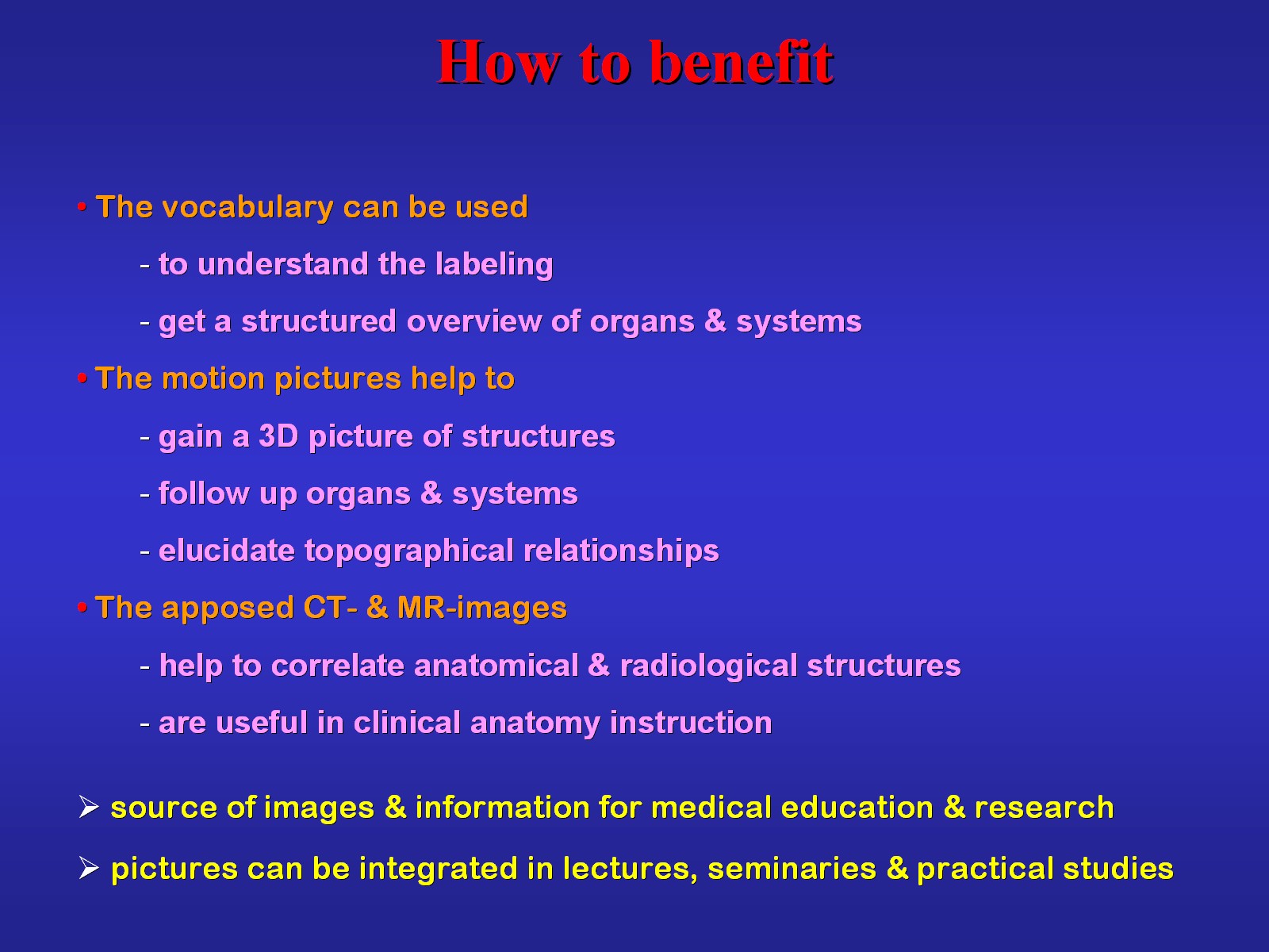 |
Fig.35
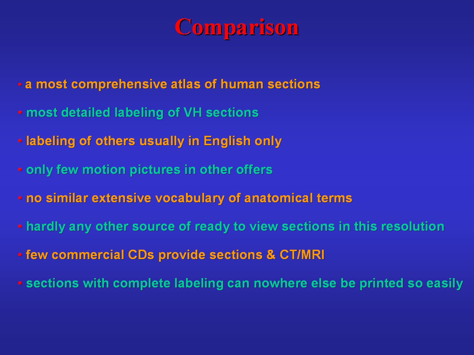 |
It is apparent that the present atlas is highly comprehensive. Elsewhere,
comparable detailed labelling, usually in English, is only present in few
commercial CDs and books. For the reasons listed in Fig.35, the
atlas represents a unique concept of presenting cross-sectional anatomy.
The atlas is constantly corrected, extended and updated. In Fig.36 you see which work is in progress. |
Fig.36
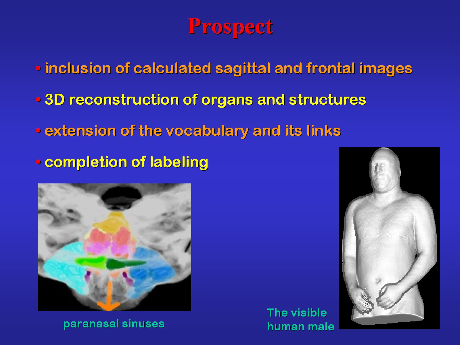 |
Fig.37
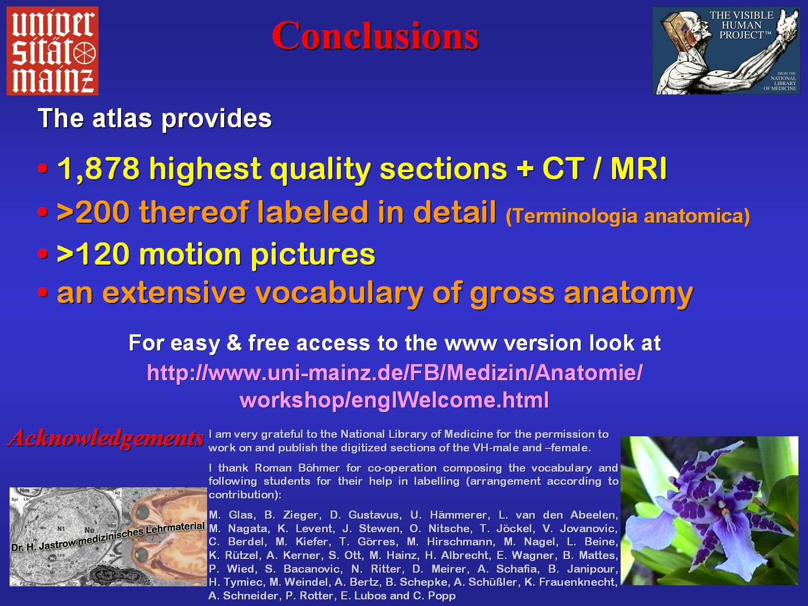 |
With the summary of Fig.37 I want to invite you to explore the pages of the workshop anatomy for the internet. The latter will include the web version of this most extensive atlas of human sectional anatomy from December on. Further, I acknowledge those who supported this project. | If you are interested in the atlas,
please contact:
specialist in anatomy Kotthaushang 22, 45239 Essen, Germany Phone: +49 201 4749893 |
--> Publications &
Presentations of Dr. Jastrow
--> Workshop Homepage
--> Imprint, Data
protection policy