Supplemental
material for the following published paper:
Analysis of synaptic bodies in the Sprague-Dawley
rat pineal gland under extreme photoperiods
Holger Jastrow and Jörg Racke
Micron
Volume 38, Issue 3: 237-251 (April 2007).
Corresponding author: PD Dr. med. Holger
Jastrow
E-mail: info@drjastrow.de
To start the animations click on the images,
please!
Color: green
= synaptic bodies
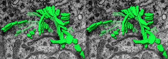
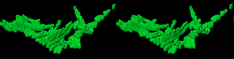
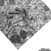
A. different stereo motion
pictures of a reconstructed field of synaptic bodies inside a Sprague-Dawley
rat pinealocyte with a section as background
The animations demonstrate the three-dimensional
morphology of 7 synaptic bodies (sbs). They all are ribbons, some are strongly
bent, one seems to have resulted from two
fused sbs. In order not to show the structures
as closely as possible to their true shape no interpolations or smoothing
algorithms were applied. Two sbs look somewhat
disrupted due to fact that they were nearly sagittally
cut in the original sections. All sbs are more or less bent in at least
one direction and only two are completely contained in
the investigated tissue volume. Also note the
deep invagination of the cell membrane close to the field and several clathrin
coated vesicles. The oriented stack of original images
from which the reconstructions were done is shown
in C.
The reconstructed field
rotates along the X-axis
(file size: 11.8 MB!) |
 |
The reconstructed field
is seen from above and
rotates along the Y-axis
(file size: 11.8 MB!) |
 |
The reconstructed field
is regarded from below and
rotates along the Y-axis
(file size: 11.8 MB!) |
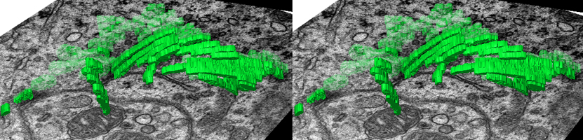 |
The reconstructed field
is visible from an oblique angle
and rotates along the Y-axis
(file size: 10.4 MB!) |
 |
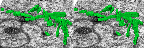
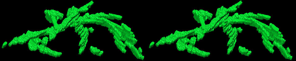
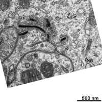
B. stereo animations demonstrating
reconstructed synaptic bodies of the same field without background
The reconstructed
field
is seen from above
and rotates along the Y-axis
(file size: 8.6 MB!) |
 |
The reconstructed field
is regarded from above
at a different angle
and rotates along the Y-axis
(file size: 8.6 MB!) |
 |
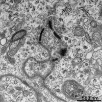
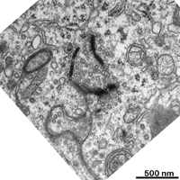
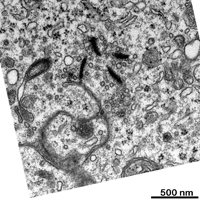
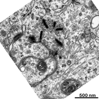
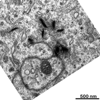
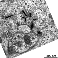
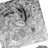
C. oriented stack of original
micrographs used for reconstruction of the field visualized in A
and B
 |
 |
 |
 |
 |
 |
|
1
|
2
|
3
|
4
|
5
|
6
|
 |
 |
 |
 |
 |
 |
|
7
|
8
|
9
|
10
|
11 (interpolated, original
damaged)
|
12
|
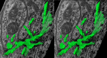
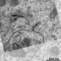
D. stereo animation in 3D "true colour", i.e.
original grey tone as in the micrographs, from another reconstructed area
| Three plate-like SBs close to the
membranes of two adjacent cells one of which clearly shows the pentalamellar
structure of SBs that is only visible when they are exactly seen from the
direction of their long axis, i.e., if they are positioned exactly at right
angle to the section plane. |
 |
--> further information on: synaptic
bodies, pineal gland
--> electron microscopic atlas homepage
--> workshop homepage