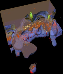
red = immunolabelling
blue = cell surface
gray = cell interior
green = synaptic ribbons
(file size: 14.4 MB!)
Holger Jastrow*, Peter Koulen# , Wilko D. Altrock°,
and Stephan Kröger* §
* Dept. of Anatomy and Cell Biology, University
of Mainz, Becherweg 13, D-55128 Mainz, Germany
# Dept. of Pharmacology & Neuroscience,
3500 Camp Bowie Blvd., 76107-2699 Fort Worth, USA
° Leibniz Institute for Neurobiology,
Brenneckestr. 6, D-39118 Magdeburg, Germany
§ Dept. of Physiological Chemistry, University
of Mainz, Duesbergweg 6, D-55099 Mainz, Germany
Author for correspondence: S. Kröger
at University of Mainz
Tel: ++49-6131-3925797
FAX: ++49-6131-3920136
email: skroeger@uni-mainz.de
Please note that the above information is
from the year of publication!
Prof. S. Kröger now works at the LMU in
Munich, Germany
A. motion pictures showing reconstructed structures turning 360° immunolabelling in red
 |
Colors:
red = immunolabelling blue = cell surface gray = cell interior green = synaptic ribbons (file size: 14.4 MB!) |
B. motion pictures showing reconstructed structures
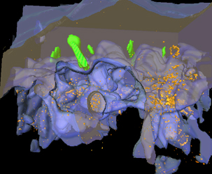
turning 360° immunolabelling in orange
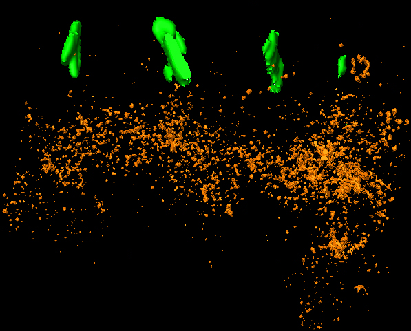
The left animation shows immunolabelling and
ribbons in the cone terminal, which is omitted in the right motion picture
 |
 |
Colors:
orange = immunolabelling blue = cell surface gray = cell interior green = synaptic ribbons (file sizes: ~11,9 MB!) for stereo animation
|
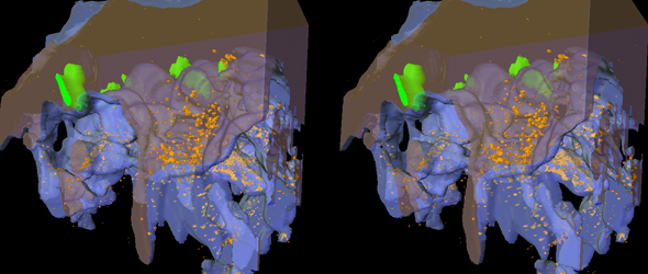
C. stereo animations of a cone synaptic terminal in blue
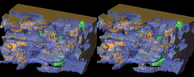
angular view |
up and downside view |
| Colors:
orange = immunolabelling; blue = cell surface; gray = cell interior; green = synaptic ribbons; (file sizes ~11.6 MB!) |
D. stereo motion pictures of a cone synaptic terminal in gray
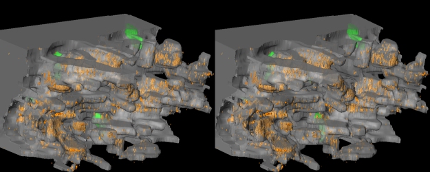
angular view |
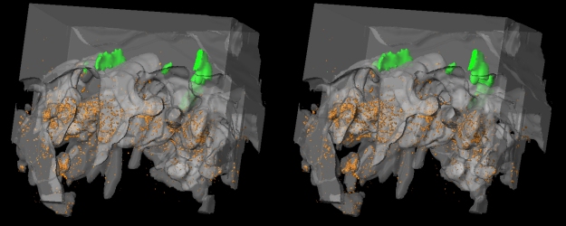
up and downside view |
| Colors:
orange = immunolabelling; light gray = cell surface; dark gray = cell interior; green = synaptic ribbons; (file sizes 11.3 - 11.6 MB!) |
E. stereo animation of synaptic ribbons and immunolabelling only
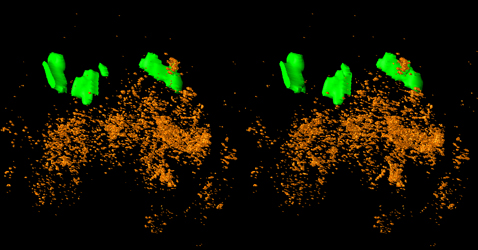 |
Colors:
orange = immunolabelling; green = synaptic ribbons (file size: 14.4 MB!) |